理解cfDNA和ctDNA在癌症生物学与诊断中的关键生物标志物
什么是细胞游离DNA(cfDNA)?
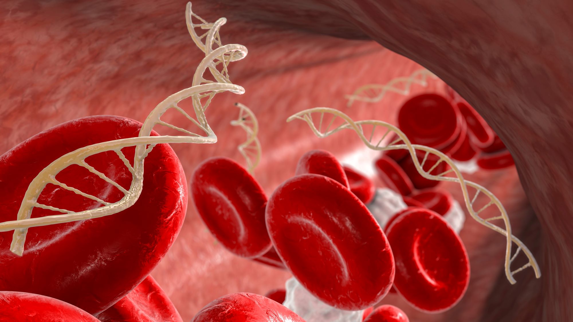
在全血的细胞游离部分及其他体液中可以发现片段化的DNA。这些被称为细胞游离DNA(cfDNA)的核酸,其大小各异(约40-1000个碱基对(bp)),但平均约为166 bp。1cfDNA由正常细胞过程以及死亡和凋亡的细胞释放到血浆中,半衰期相对较短,大约为5-150分钟。2在健康个体中,血浆DNA水平通常低于10 ng/mL,但在某些特定疾病状态下,如心肌梗死、中风和糖尿病,可显著升高。3,4鉴于cfDNA具有瞬时性和动态变化的特点,并且会在特定疾病状态下升高,因此cfDNA可作为一种生物标志物,用于检测、分期和评估多种不同的健康状况和疾病状态。
cfDNA可以从多种不同的生物流体中提取,包括血浆或血清、脑脊液、尿液、唾液以及胸腔积液。5 这种被称为“液体活检”的方法,比放射学检查和组织活检更经济且创伤性更小。2,6,7因此,与通过常规途径获取生物样本相比,在多项临床研究和应用中,利用cfDNA作为诊断标志物已被证明是可行的。cfDNA的使用已被证实适用于孕妇的产前诊断目的。8–10此外,已有研究表明,cfDNA可用于人类器官移植中早期检测排斥反应。11在新冠疫情期间,cfDNA被用来评估新冠患者的组织损伤程度。12最后,目前cfDNA中的甲基化模式已被用于大规模结直肠癌筛查。13
循环肿瘤DNA(ctDNA),cfDNA的一种亚型
鉴于cfDNA能够代表潜在生物过程的“快照”,无论是细胞群体还是细胞功能发生显著变化,都有望在可检测的cfDNA含量和丰度中得到反映。在癌症的特定背景下,快速的克隆扩增和选择压力将推动疾病进展,同时增加循环肿瘤DNA(ctDNA)的水平。14ctDNA被认为是cfDNA的一个子集,通常比非突变型cfDNA分子更短。7近年来,由于所用生物标志物具有无与伦比的特异性,ctDNA的检测在癌症诊断领域获得了极大的关注。这些肿瘤特异性生物标志物主要是指在癌前或癌细胞中检测到的DNA突变,最常见的是单碱基对替换。
由于这种无与伦比的肿瘤生物标志物特异性,对ctDNA作为诊断、预后及研究主题的兴趣激增。尽管这一领域仍处于发展阶段,对ctDNA真正价值的理解还在不断深入,但迄今为止已有证据支持这样一种假设:ctDNA就像“火灾前冒出的烟”。在早期诊断方面,研究表明,在转移性疾病出现之前,检测到ctDNA对于更早发现癌症具有重要意义。事实上,有研究已在实际确诊癌症前长达2年,在唾液和血浆中检测到了相关突变。15对于正在进行分期和治疗中的癌症患者,研究发现ctDNA的检测结果与肿瘤大小和分期均呈相关性。16,17此外,与其他常用肿瘤标志物相比,ctDNA显示出更高的预后能力。7当原发肿瘤来源不明时,可以利用特定甲基化及核小体占据模式来编码组织或细胞特异性的信息。18
除了癌症的诊断和分期之外,通过获取对恶性肿瘤的整体视角,ctDNA的利用还可以根据特定突变的有无,更好地为个体化治疗方案提供信息。传统活检方法可能因局部取样而低估或引入偏差,从而影响个体化药物的选择和疗效。事实上,多区域测序研究已经证明,同一患者不同肿瘤区域的突变谱存在异质性。19,20因此,通过液体活检获得、来源于多个肿瘤区域的ctDNA分析,可能更能反映整体肿瘤异质性,并防止通过单一区域活检取样所带来的偏倚。
最后,ctDNA还可以在治疗期间及治疗后进行纵向监测。多项研究已将ctDNA动态变化与治疗反应相关联。7与其他肿瘤标志物相比,ctDNA监测不仅能最早检测到可测量的治疗反应,还表现出最大的动态范围,并且是复发最早期的指示信号。21此外,监测ctDNA还能揭示克隆进化以及当前治疗方案耐药性的产生。对肿瘤突变谱进行多重检测,可以发现相对变化,并为了解肿瘤分子进化提供线索。22,23
通过精准提取和分离,实现全面cfDNA和ctDNA检测
鉴于使用ctDNA作为诊断和预后指标的诸多益处,这种方法已经获得了显著的发展。然而,ctDNA检测以及与正常cfDNA的区分一直是技术上的挑战。目标突变序列的存在,尤其是在疾病早期或治疗后肿瘤负荷较低的情况下,与整体cfDNA含量相比,其拷贝数极低。6实际上,在疾病早期阶段,ctDNA仅占整体cfDNA含量的不到1%。24,25然而,核酸扩增技术在检测和分辨能力方面的同步提升,使得这些极低拷贝数事件得以被检测到。新型数字PCR方法结合下一代测序技术,由于增强了对稀有目标序列的检测能力以及多个相关基因的多重检测,使得对ctDNA的深入研究成为可能。21,26
尽管基因突变检测方法有了极大改进,但样本制备同样重要。分离DNA是cfDNA检测流程及从正常cfDNA中区分ctDNA的重要环节。样本纯化工具如MagMAX试剂盒以及KingFisher仪器对于利用cfDNA深入理解癌症致病机制,以及推动癌症早期诊断的发展都至关重要。Applied BiosystemsTM MagMAXTM 无细胞DNA分离试剂盒旨在富集循环cfDNA,并针对血清和血浆等生物样品进行了优化。该技术采用磁珠技术,能够稳定回收高质量的DNA,适用于实时PCR、数字PCR和下一代测序等广泛应用。同样,KingFisher仪器是极其多功能且自动化的纯化系统,可为低通量和高通量工作流程提供高效、可重复且可靠的结果。最后,赛默飞还提供了丰富的信息资源,涵盖最新的生物标志物研究以协助方法开发的各个方面。结合更高通量的样品制备与更敏感的扩增方法,共同推动基础癌症研究和诊断开发实现癌症更早期检测及更全面治疗策略。
仅供科研使用。不得用于诊断程序。
##
参考文献
1. Kustanovich, A., Schwartz, R., Peretz, T. & Grinshpun, A. Life and death of circulating cell-free DNA. Cancer Biology and Therapy vol. 20 1057–1067 Preprint at https://doi.org/10.1080/15384047.2019.1598759 (2019).
2. Song, P. et al. Limitations and opportunities of technologies for the analysis of cell-free DNA in cancer diagnostics. Nature Biomedical Engineering vol. 6 232–245 Preprint at https://doi.org/10.1038/s41551-021-00837-3 (2022).
3. Luo, H., Wei, W., Ye, Z., Zheng, J. & Xu, R. hua. Liquid Biopsy of Methylation Biomarkers in Cell-Free DNA. Trends in Molecular Medicine vol. 27 482–500 Preprint at https://doi.org/10.1016/j.molmed.2020.12.011 (2021).
4. Gaitsch, H., Franklin, R. J. M. & Reich, D. S. Cell-free DNA-based liquid biopsies in neurology. Brain vol. 146 1758–1774 Preprint at https://doi.org/10.1093/brain/awac438 (2023).
5. Ponti, G., Manfredini, M. & Tomasi, A. Non-blood sources of cell-free DNA for cancer molecular profiling in clinical pathology and oncology. Critical Reviews in Oncology/Hematology vol. 141 36–42 Preprint at https://doi.org/10.1016/j.critrevonc.2019.06.005 (2019).
6. Diaz, L. A. & Bardelli, A. Liquid biopsies: Genotyping circulating tumor DNA. Journal of Clinical Oncology vol. 32 579–586 Preprint at https://doi.org/10.1200/JCO.2012.45.2011 (2014).
7. Wan, J. C. M. et al. Liquid biopsies come of age: Towards implementation of circulating tumour DNA. Nature Reviews Cancer vol. 17 223–238 Preprint at https://doi.org/10.1038/nrc.2017.7 (2017).
8. Dennis Lo, Y. M. et al. Presence of Fetal DNA in Maternal Plasma and Serum. THE LANCET vol. 350 (1997).
9. Lo, Y. M. D. & Chiu, R. W. K. Genomic analysis of fetal nucleic acids in maternal blood. Annual Review of Genomics and Human Genetics vol. 13 285–306 Preprint at https://doi.org/10.1146/annurev-genom-090711-163806 (2012).
10. Ennis, Y. M. D. et al. PRENATAL DIAGNOSIS OF FETAL RhD STATUS BY MOLECULAR ANALYSIS OF MATERNAL PLASMA A BSTRACT Background The Ability to Determine Fetal RhD. (1998).
11. De Vlaminck, I. et al. Noninvasive monitoring of infection and rejection after lung transplantation. Proc Natl Acad Sci USA 112, 13336–13341 (2015).
12. Andargie, T. E. et al. C L I N I C A L M E D I C I N E Cell-free DNA maps COVID-19 tissue injury and risk of death and can cause tissue injury. (2021) doi:10.1172/jci.
13. Lamb, Y. N. & Dhillon, S. Epi proColon® 2.0 CE: A Blood-Based Screening Test for Colorectal Cancer. Mol Diagn Ther 21, 225–232 (2017).
14. Kasimir-Bauer, S. et al. Does primary neoadjuvant systemic therapy eradicate minimal residual disease? Analysis of disseminated and circulating tumor cells before and after therapy. Breast Cancer Research 18, (2016).
15. Gormally, E. et al. TP53 and KRAS2 mutations in plasma DNA of healthy subjects and subsequent cancer occurrence: A prospective study. Cancer Res 66, 6871–6876 (2006).
16. Kamat, A. A. et al. Circulating cell-free DNA: A novel biomarker for response to therapy in ovarian carcinoma. Cancer Biol Ther 5, 1369–1374 (2006).
17. Bettegowda, C. et al. Detection of circulating tumor DNA in early- and late-stage human malignancies. Sci Transl Med 6, (2014).
18. Rosenfeld, N. et al. MicroRNAs accurately identify cancer tissue origin. Nat Biotechnol 26, 462–469 (2008).
19. Yates, L. R. et al. Subclonal diversification of primary breast cancer revealed by multiregion sequencing. Nat Med 21, 751–759 (2015).
20. Gerlinger, M. et al. Intratumor Heterogeneity and Branched Evolution Revealed by Multiregion Sequencing. New England Journal of Medicine 366, 883–892 (2012).
21. Dawson, S.-J. et al. Analysis of Circulating Tumor DNA to Monitor Metastatic Breast Cancer. New England Journal of Medicine 368, 1199–1209 (2013).
22. Diaz, L. A. et al. The molecular evolution of acquired resistance to targeted EGFR blockade in colorectal cancers. Nature 486, 537–540 (2012).
23. Misale, S. et al. Emergence of KRAS mutations and acquired resistance to anti-EGFR therapy in colorectal cancer. Nature 486, 532–536 (2012).
24. Diehl, F. et al. Detection and Quantification of Mutations in the Plasma of Patients with Colorectal Tumors. www.pnas.orgcgidoi10.1073pnas.0507904102 (2005).
25. Holdhoff, M., Schmidt, K., Donehower, R. & Diaz, L. A. Analysis of circulating tumor DNA to confirm somatic KRAS mutations. Journal of the National Cancer Institute vol. 101 1284–1285 Preprint at https://doi.org/10.1093/jnci/djp240 (2009).
26. Murtaza, M. et al. Non-invasive analysis of acquired resistance to cancer therapy by sequencing of plasma DNA. Nature 497, 108–112 (2013).

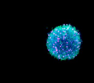
癌症类器官培养技巧
癌症类器官是从患者肿瘤样本中获得的三维细... Bioprocessing Staff
了解更多
siRNA与miRNA:解码这些微小却强大的分子
RNA干扰(RNAi)是一种高度保守且至关重要的�... Dana D'Amico
了解更多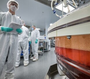
细胞治疗制造中的原材料考量
在细胞治疗制造过程中,缺乏明确的监管框架... Tanuka Biswas, PhD
了解更多
显微图像去卷积:清晰显微镜指南
显微镜极大地提升了我们观察不可见世界的能... Dana D'Amico
了解更多
发表回复