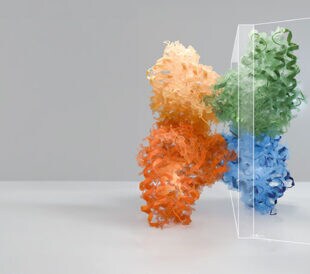
Over the last year, researchers turned to cryo-electron microscopy (cryo-EM) to increase the human understanding of the SARS-CoV-2 virus, as they have done with other major viruses like Zika, Ebola, and Human Immunodeficiency Virus. Our cryo-EM instruments, including the Krios Cryo-TEM, have been used for breakthrough coronavirus research findings, such as one recently published by a team of researchers at Heidelberg University.
Coronavirus Research Fueled by Powerful Imaging
In 2020, this team quickly set out to understand how the virus changes infected cells to achieve effective reproduction and made significant headway by characterizing the replication of SARS-CoV-2 inside host cells.
Using cryo electron tomography (cryo-ET), a cryo-electron microscopy technique that provides three-dimensional images of molecular structures at nanometer resolution, the researchers revealed how SARS-CoV-2 changes the cellular membranes and organelles of infected cells, which is critical for effective replication.

The researchers performed molecular analyses of the viral RNA and individual steps of virus replication to obtain unprecedented detail of the network composed of double-stranded RNA inside the double-membrane vesicles.
Cryo-Electron Tomography of Infected Cells
Using in-situ cryo-ET to image 200-nanometer thin layers of virus-infected cells, the researchers discovered how the viral RNA creates the necessary factors for its replication, allowing it to quickly reproduce and possibly even protect itself against immune system attacks. Using our Thermo Scientific Krios Cryo-TEM and Aquilos Cryo-FIB in combination with Thermo Scientific Amira Software, they rendered the three-dimensional environment of the cell—while visualizing individual virus particles and details of the virus assembly sites.
These discoveries built on the team’s previous experience studying viral infections such as Influenza A and Ebola. “Thanks to our expertise in cryo-electron tomography, the excellent microscopes at the campus of Heidelberg University, and the highly collaborative environment at the Center for Integrative Infectious Disease Research, we were able to start detailed molecular examinations on SARS-CoV-2 infected cells right at the beginning of the coronavirus pandemic,” said Heidelberg University Hospital researcher Dr. Petr Chlanda.
The researchers’ next step with their coronavirus research is to uncover which proteins are involved in the formation of the double membrane structures. Characterizing the replication cycle of SARS-CoV-2 inside the host cell is key to developing drugs that restrict their reproduction—and the Heidelberg University researchers have brought us a big step closer to that goal.
Read our customer spotlight to learn more.
//
Séverine Baillet, PhD, is a marketing project manager at Thermo Fisher Scientific.
Speak with an expert: https://www.thermofisher.cn/blog/microscopy/speak-with-an-expert/
Subscribe now to receive Accelerating Microscopy updates straight to your inbox.
Advances in high-resolution cryo-EM at 100 kV
100 kV cryo-TEM enables high-resolution single particle anal... Alex Ilitchev, PhD
Read More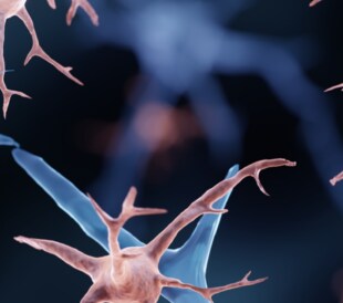
3D Tissue Histology with Light-Sheet Microscopy Enables Nondestructive Analysis of Microglia
3D tissue analysis offers critical benefits for neuroscience... Alex Ilitchev, PhD
Read More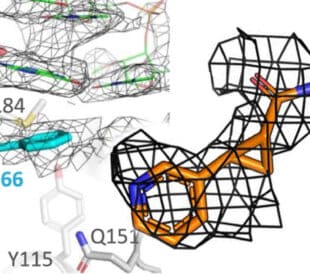
Fragment based drug discovery meets challenging drug targets with high-throughput cryo-EM
Benefits of FBDD in the search for novel therapeutics Frag... Dominic Meusch
Read More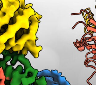
Targeted protein degradation as a novel therapeutic approach for undruggable diseases
Induced proximity for targeted protein degradation In 1993, ... Dominic Meusch
Read More
Leave a Reply