Scientists at academic institutions continue to work on the cutting-edge of cryo-EM research. To celebrate the work these scientists are doing to solve some of the world’s most complex problems, we’re hosting a “Cryo-EM in the Classroom” series to spotlight the latest research in academia.
Using cryo-electron microscopy (cryo-EM), today’s scientists aren’t limited to low-resolution 2D images but instead have the ability to image high-resolution 3D structures. By studying these 3D models, researchers gain a better understanding of the structure and interactions of certain proteins at the atomic level — the same level of resolution that physicists use to study beams of light. Ultimately, these new insights can speed the path to more effective treatments against viruses and diseases.

Detailed reconstructions are now available with modern cryo-EM. 5N8Y. KaiCBA circadian clock backbone model based on a Cryo-EM density via DOI: 10.1126/science.aag3218
For example, Dr. Timothy Baker from the University of San Diego Division of Biological Sciences has applied cryo-EM in his studies for over two decades. Dr. Baker uses cryo-EM instruments to create 3D structures to better understand the interactions a virus has with a host cell as it replicates. In one such study, Dr. Baker used cryo-EM to visualize how different proteins latch onto a bacterial host, and how the tail spike protein changes conformations to facilitate infection as shown here.
Just this year, researchers at Purdue University leveraged cryo-EM to map out the complexities of the Zika virus and show the structure of a mature Zika virus at 3.1Å resolution. Two years prior, those same researchers were only able to produce a resolution of 3.8Å. With new virus preparation methods and updated data processing techniques, the team was able to refine the quality of their structures. Access to this detailed 3D structure of the Zika virus equips researchers with crucial information in their move to design more effective and targeted vaccines.

Cryo-EM structure of mature Zika virus at 3.1Å resolution. (Purdue University photo/Madhumati Sevvana)
Alzheimer’s is a neurological epidemic that has been studied for over a century. Over time, and often later in life, the neurodegenerative disease causes memory loss and impaired thinking, eventually leading to the inability to carry out simple tasks. In the late 1980s, scientists learned that tau protein was a key component of the disease, but it wasn’t until 2017 that a team of researchers, led by Dr. Sjors Scheres and Dr. Michel Goedert of MRC Laboratory of Molecular Biology, visualized the filaments’ atomic structure. Dr. Anthony Fitzpatrick, who participated in the 2017 research, and his group at Columbia University’s Zuckerman Institute continue to bring new voices to the table to decipher the disease, in hopes that their research will lay foundations for future breakthroughs.

Tau filaments as imaged with cryo-EM. Credit: Anthony Fitzpatrick/Columbia’s Zuckerman Institute
Stay tuned for more posts in this Cryo-EM in the Classroom series where we’ll profile amazing cryo-EM work from researchers at academic and research institutions around the globe. In the meantime, check out EM-Learning.com, an online learning center where you can access more than 50 hours of lectures and videos on all things cryo-EM – at no cost to you!
Do you know someone who should be featured for their cryo-EM work? Reach out to us on Twitter or LinkedIn. Subscribe to receive Accelerating Microscopy updates straight to your inbox.
To learn more about cryo-EM, fill out this form to speak with an expert.

Advances in high-resolution cryo-EM at 100 kV
100 kV cryo-TEM enables high-resolution single particle anal... Alex Ilitchev, PhD
Read More
3D Tissue Histology with Light-Sheet Microscopy Enables Nondestructive Analysis of Microglia
3D tissue analysis offers critical benefits for neuroscience... Alex Ilitchev, PhD
Read More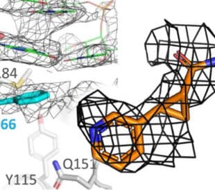
Fragment based drug discovery meets challenging drug targets with high-throughput cryo-EM
Benefits of FBDD in the search for novel therapeutics Frag... Dominic Meusch
Read More
Targeted protein degradation as a novel therapeutic approach for undruggable diseases
Induced proximity for targeted protein degradation In 1993, ... Dominic Meusch
Read More
Leave a Reply