Glacios 2 Cryo-TEM and the Evolution of 200 kV Cryo-EM
Cryo-electron microscopy (cryo-EM) has come a long way since the first viral structures were published by the Dubochet group in the early 1980s. Year over year, there is an almost exponential increase in the number of cryo-EM structures submitted to the Protein Data Bank (PDB), many at resolutions better than 3 Å. Notably, these structures are often for membrane proteins and other systems that have been resistant to traditional crystallographic analysis.
This advancement was made possible by the evolution of cryo-EM technology, including the new 200 kV Thermo Scientific Glacios 2 Cryo-TEM, which delivered early and impressive results after it was announced in October 2022. In March 2024, Themo Fisher Scientific introduced an optional low-energy-spread cold field emission gun (E-CFEG) that, in combination with the Glacios 2 Cryo-TEM, broke the world record for resolution at 200 kV for apoferritin (1.5 Å).
Realizing potential with the Glacios 2 Cryo-TEM
The vast majority of sub-3 Å structures submitted to the PDB were determined with 300 kV cryo-transmission electron microscopes (cryo-TEMs) that were specifically designed for high-resolution data collection at cryogenic temperatures. In fact, it is safe to say that 300 kV has been treated as the de facto accelerating voltage for high-quality structural analysis. When they were introduced, 200 kV instruments such as the Thermo Scientific Talos Arctica and Glacios Cryo-TEMs were, therefore, largely seen as screening tools that could be used to optimize samples without spending time on the higher-resolution 300 kV instruments.
However, advanced components and software are not reserved exclusively for 300 kV microscopes. They have allowed 200 kV cryo-TEMs to experience their own “resolution revolution,” and research groups around the world are beginning to produce highly valuable results directly at 200 kV.

Prof. Cristina Paulino formerly at the University of Groningen highlighted her previous group’s development of an optimized cryo-EM data-acquisition workflow by sample-thickness determination. Learn more >>
A revolution in productivity and cryo-EM resolution
The new Glacios 2 Cryo-TEM showcases many of the advancements that have made high-quality analysis possible on 200 kV instruments. Hardware optimization has focused on improved specificity and sensitivity in electron detection. For instance, the Thermo Scientific Falcon 4i Direct Electron Detector enables high detective quantum efficiency (DQE) across the full spectral range, capturing both small and flexible proteins as well as larger structures using fewer images. When coupled with a novel Thermo Scientific Selectris or Selectris X Energy Filter, signal is limited to the zero-loss, elastically scattered electrons, producing high-contrast TEM data for higher throughput and higher resolution structures. In addition, the E-CFEG option provides a narrow energy spread that results in higher-contrast images and higher resolution. This add-on makes it possible to efficiently solve structures at resolutions of 2 Å or less.
With the introduction of Thermo Scientific Smart EPU Software, both automated screening and data acquisition for single particle cryo-EM analysis are now available through an approachable graphical user interface. Featuring unique AI algorithms, Smart EPU Software allows for thousands of images to be collected reproducibly and with minimal user input, and integrates aberration-free image shift (AFIS), producing more accurate, usable data with each scan.

A reconstruction of apoferritin at 1.5 Å resolution from data collected over eleven hours using the Glacios 2 Cryo-TEM with the Falcon 4i Direct Electron Detector, Selectris Imaging Filter, E-CFEG, and FFI.
What follows are just a few examples of how 200 kV cryo-TEM is being utilized by researchers internationally to produce novel, actionable results.

Prof. Gabe Lander’s group at the Scripps Research Institute published their 2.7 Å reconstruction of transthyretin (55 kDa) using 200 kV cryo-TEM. Cryo-EM reveals new information that explains amyloidogenic pathway.
Drug discovery at 200 kV
Structure-based drug design has been a longstanding goal of pharmaceutical research, as it enables the development of targeted treatments based on molecular insights. Efforts have largely been hampered by the speed at which actionable data can be collected and processed. Modern cryo-EM, however, has made significant strides toward structure-enabled therapeutics thanks to high-throughput, high-resolution data.
For example, in March 2024, Professor Basil Greber of the Institute of Cancer Research, London, published a paper in Nature Communications demonstrating how the Glacios 2 Cryo-TEM was used to determine the structure of a CDK-activating kinase (CAK). These enzymes are involved in cell regulation and are actively being investigated as potential targets for cancer treatments.

CDK-activating kinase (CAK) complex structures solved on a Glacios 2 Cryo-TEM. Drug binding occupancy on this complex was analyzed following a fast-screening strategy of only a few hours of data collection per sample. A large dataset enabled a 2.3 Å structure in less than a day where an atomic model could be built unambiguously. This project was a collaboration with Basil Greber from Institute of Cancer Research, London, and Professors Simak Ali and Matthew J. Fuchter from Imperial College, London.
Read application note: Powering drug discovery with one structure per hour using cryo-EM>
GPCR structure determination
G protein-coupled receptors (GPCRs) compose a large class of membrane proteins that are promising therapeutic targets for a variety of diseases, but they are notoriously difficult to crystallize, hampering crystallographic characterization. Cryo-EM has made significant headway in the analysis of GPCRs in recent years, but this work has been limited to 300 kV instruments. However, researchers at Monash University were able to successfully determine the structure of a GPCR to 3.2 Å resolution on a 200 kV Glacios Cryo-TEM, a platform whose general ease of use and increased throughput has the potential to significantly broaden structure-based drug design for GPCRs.
μ-Opioid Receptor cryo-EM structure with nanobody antagonists
The μ-opioid receptor (μOR) is a prototypical member of the G protein-coupled receptor (GPCR) family. Due to the recent opioid crisis, there is an interest in developing new modulators of μOR function. Andreas Borland’s lab, using a Talos Arctica Cryo-TEM in combination with a Falcon 3EC Detector, determined the structure of μOR with the nanobody NbE (NbE-µOR complex) to 3.5 Å. The structure explains the high selectivity of NbE and provides insights for further drug design targeting μOR.

Cryo-EM structure of NbE-µOR complex and close up of the NbE-µOR interaction interface.
Cryo-EM of human fucosidase

Close up of the FucA1 catalytic center without (A) and with (B) a bonded ligand. (C) shows several bacterial fucosidases overlaid onto the structure of FucA1. Figure reproduced from the original article by Zachary Armstrong et al under CC BY 4.0.
The enzyme fucosidase plays a vital role in certain bacterial infections as well as fucosidosis, a neurodegenerative disorder impacting lysosomal storage. A complete understanding of fucosidase catalysis, along with any potential targeted drug design, has been hampered by the lack of 3D structures for the enzyme. Utilizing a Glacios Cryo-TEM, researchers at the University of York were able to resolve the structure of human lysosomal α-L-fucosidase (FucA1) down to 2.49 Å resolution. They were also able to clearly visualize FucA1 in complex with an inhibitor, deoxyfuconojirimycin. At this level of detail, the precise architecture of the catalytic center could be determined as well as the location of various disease-related mutations. This serves as a promising step toward targeted treatment of fucosidase-related diseases and disorders.

Jamie Blaza, a contributor on the article, shares some of the beautiful 2D classes they obtained for FucA1.
200 kV advancements beyond single particle cryo-EM: MicroED of organometallics
Microcrystal electron diffraction (microED) is a burgeoning small molecule characterization technique propelled by the increased availability of high-resolution cryo-electron microscopy equipment. MicroED can analyze much smaller samples (i.e., nanocrystals) compared to X-ray diffraction, and, unlike other electron diffraction techniques, is designed to be compatible with sensitive organic samples such as protein crystals.
A recent study from the University of York showcases how 200 kV cryo-TEM can be used for the MicroED analysis of single crystal organometallics. Specifically, single crystal to single crystal (SC-SC) transformations, performed on grid, can be used to generate and characterize otherwise highly reactive organometallic species. Here, nanocrystals composed of a norbornadiene complex were hydrogenated by the addition of H2 gas into a σ-alkane (norbornane) complex. The resulting crystallographic structures were resolved down to 0.95 Å resolution. These results not only showcase the quality of MicroED data that can be generated at 200 kV, but more broadly demonstrate the potential of cryo-EM for on-grid single-crystal chemistry.

Comparison of MicroED data collected from a nanocrystal (C) with traditional X-ray diffraction results obtained on a larger crystal that was much more difficult to obtain. Reproduced from the corresponding article by LR Doyle et al under CC BY 3.0.
Cryo-electron tomography on the Glacios 2 Cryo-TEM
Tomography is a reconstructive imaging technique that combines multiple 2D snapshots collected at different angles into a single 3D dataset. Cryo-electron tomography (cryo-ET), specifically, is used on thinned cell samples to obtain high-resolution 3D information on organelles and proteins within their cellular context. Using the Glacios 2 Cryo-TEM, tomography data can be obtained from whole bacterial cells or lamellae prepared with Thermo Scientific Aquilos or new Arctis Cryo-Focused Ion Beam (Cryo-FIB) instruments. In the Magnetospirillum example shown below, membranous compartments, filaments, and larger proteins can clearly be visualized within the 300 nm thick plunge-frozen bacterial cell.

3D visualization of the cell membrane, liposomes, and filaments in a Magnetospirillum bacterium, generated using 200 kV Glacios 2 Cryo-TEM data. Sample courtesy of Dirk Schüler, University of Bayreuth.
The future of 200 kV cryo-EM
Cryo-EM software and hardware continue to improve at an astonishing pace, painting a bright future for the technique. Notably, cutting-edge data processing, along with AI-driven modeling of cryo-EM data, is showing incredible potential for taking us from sample to structure faster than ever before.

Martin Pacesa of EPFL shows the development of a 3.3 Å reconstruction in under an hour using the novel Model Angelo modeling tool.

Advances in high-resolution cryo-EM at 100 kV
100 kV cryo-TEM enables high-resolution single particle anal... Alex Ilitchev, PhD
Read More
3D Tissue Histology with Light-Sheet Microscopy Enables Nondestructive Analysis of Microglia
3D tissue analysis offers critical benefits for neuroscience... Alex Ilitchev, PhD
Read More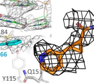
Fragment based drug discovery meets challenging drug targets with high-throughput cryo-EM
Benefits of FBDD in the search for novel therapeutics Frag... Dominic Meusch
Read More
Targeted protein degradation as a novel therapeutic approach for undruggable diseases
Induced proximity for targeted protein degradation In 1993, ... Dominic Meusch
Read More
Leave a Reply