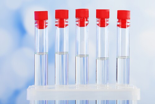
 Because cerebrospinal fluid (CSF) is adjacent to and interacts with the brain’s parenchyma, it yields brain-specific molecules reflective of the pathophysiology of neurological diseases and brain-based disorders. These include bacterial meningitis, multiple sclerosis and Alzheimer’s disease. For this reason, CSF is particularly valuable for analysis, including biomarker discovery. The identification of novel CSF biomarkers could allow for early diagnosis, prognostic prediction, disease monitoring and the development of neuroprotective treatments.
Since the techniques for obtaining even lumbar CSF samples are invasive, samples from healthy controls are rare, and few large CSF biobanks exist. This means that scientific collaboration for access to this resource is at a premium, requiring standardized sampling and storage protocols. In a recent review, Willemse and Teunissen (2015) indicate that enhanced industry understanding of the value of CSF biobanking and strategies to address long-term storage quality control challenges could reap scientific rewards.1
According to the authors, pre-analytical errors make up 60% of total laboratory errors and represent a significant issue for CSF biomarker analysis. This preanalytical variation includes factors related to the patient/donor as well as differences in collection techniques and assay performance. Additionally, the specific cellular and biochemical composition of CSF as compared with blood (e.g., lower protein concentration and protease activity) can impact protein stability. Since approximately 85% of CSF proteins derive from blood, contamination with blood can also affect biobanked samples.
Pre-Analytical Variation Factors1
|
Patient-derived |
Laboratory processing |
Assay performance |
|
Diet |
Transport time and temperature |
Buffer composition |
|
Exercise |
Time delay to spinning |
Machine settings |
|
Diurnal rhythm |
Spinning conditions |
Compliance with protocol |
|
Clinical history documentation |
Time to freezing |
Lack of certified reference materials |
|
Sample labeling |
Freezing temperature |
|
|
Duration of freezing |
||
|
Type of laboratory plastics |
When it comes to patient-derived variations, the authors recommend careful documentation for long-term storage and future research purposes. This includes fasting status, caffeine intake, smoking status, alcohol use and exercise, as well as specific time of sample collection. These variabilities can also be addressed through the use of electronic patient records linked with research databases. The authors indicate that 2-D bar coding is the gold standard, facilitating automated sample picking, human error reduction and pseudonymization.
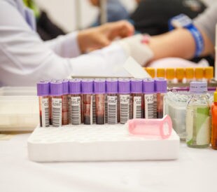
For sampling-derived variations, Willemse and Teunissen offer the following best practices for consistent collection and preparation, drawn from the consensus guideline of the BioMS-consortium:2
Collection Procedures1
|
Procedure |
Ideal Protocol for CSF |
|
Time of withdrawal and storage |
Record collection date and time |
|
Preferred volume |
At least 12 ml: 1 to 2 ml for CSF assessment and 10 ml for biobanking Record volume taken and fraction banked |
|
Location |
Intervertebral space L3-L5 (S1) |
|
If blood contamination occurred |
Do not process further Criteria for blood contimination: >500 red blood cells/μL Record number of blood cells in diagnostic samples |
|
Type of needle |
Atraumatic |
|
Type of collection tube |
|
|
Other body fluids collected simultaneously |
Serum |
|
Plasma (EDTA over citrate) |
Processing Procedures1
|
Procedure |
Ideal Protocol for CSF |
|
Storage temperature until freezing |
Room temperature before, during and after centrifugation |
|
Centrifugation conditions |
2,000 g (1,800–220), 10 minutes at room temperature |
|
Time delay between withdrawal, processing and freezing |
30 to 60 minutes, maximum 2 hours After centrifugation, aliquot and freeze samples immediately. Maximum delay 2 hours. |
|
Tubes for aliquoting |
Small polypropylene tubes (2 ml for routine diagnostics, 1 ml for biobanking) with screw caps. Record manufacturer |
|
Aliquoting |
Minimum of 2; the recommended research sample volume should provide >10 aliquots |
|
Volume of aliquots |
0.1 ml minimum, depending on total tube volume (0.2, 0.5 and 1 ml) Tubes filled up to 75% of volume |
|
Coding |
Use unique codes with freezing-proof labels. |
|
Freezing temperature |
−80 °C |
So far, researchers believe that CSF biomarkers and neurofilament proteins should be stable during long-term storage. However, there are few studies available and some indication that freezing and thawing could produce structural changes that impact CSF samples. Increasing CSF sample stores could produce experimental data useful in answering this question.
The authors also indicate that, even when facilities use the same commercial kits, they may employ different biomarker outcome levels and cut-off levels. This intra- and inter-laboratory variation has been shown to alter diagnosis in 26% and 12% of cases, respectively, specifically for the Aβ42 biomarker.3 The authors suggest that, in addition to standardizing protocols, establishing an independent quality marker for CSF would mitigate this variation. This would require the identification of a panel of sentinel molecules that are each unstable due to a specific preanalytical step and could thus highlight the source of decreased quality. Because CSF proteins are less abundant and even below the limits of detection in some cases, the identification of these markers would be challenging.
Willemse and Teunissen suggest that collaborative efforts with regard to CSF banking and research have ushered in a new era for the diagnosis and treatment of neurodegenerative diseases, particularly via biomarkers. This, combined with standardization of sampling and processing protocols, could improve biomarker research and directly impact individuals with neurodegenerative diseases.
References
1. Willemse, E. A. J. & Teunissen, C. E. (2015) “Biobanking of cerebrospinal fluid for biomarker analysis in neurological diseases,” Biobanking in the 21st Century: Advances in Experimental Medicine and Biology, 864. doi: 10.1007/978-3-319-20579-3_7
2. Teunissen, C.E., et al. (2009) “A consensus protocol for the standardization of cerebrospinal fluid collection and biobanking,” Neurology, 73 (pp. 1914–1922). doi:10.1212/WNL.0b013e3181c47cc2
3. Vos, S.J.B., et al. (2014) “Variability of CSF Alzheimer’s disease biomarkers: Implications for clinical practice,” PLoS One, 9:e100784. doi:10.1371/ journal.pone.0100784.
Engaging Diverse Populations in Biospecimen Donation
Certain subpopulations have disproportionately low participa...
Read More
Secondary Biobanking Consent in an Underrepresented Population
One of the commonly raised questions in biobanking research ...
Read More
Biobanking for HIV Malignancy Research in Sub-Saharan Africa
Sub-Saharan Africa has one of the highest incidences of HIV ...
Read More
Stabilizing Tissues for Postmortem Peptide Preservation
Classically, life stops at death; however, cellular processe...
Read More
Leave a Reply