As cryo-electron microscopy (cryo-EM) continues to mature, an increasing number of researchers from around the world use this cutting-edge technique to gain insight into many of today’s leading diseases. Scientists use cryo-EM because of its unique ability to flash-freeze samples and examine them at high resolution in their near-native state, which is leading to breakthrough discoveries. As cryo-EM instruments become easier to use while generating higher resolution results, these discoveries are taking place at an unprecedented pace.
Cryo EM advances driving novel disease research
From neurodegenerative diseases such as Alzheimer’s and Parkinson’s to infectious diseases such as COVID-19, cryo-EM is leading to new discoveries that are pushing the boundaries of research. In the last few years alone, the study of diseases using cryo-EM has led to a surge of scientific publications. Here are just a few examples:
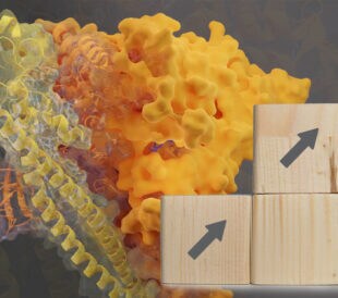
- Potential drug targets for SARS-CoV-2: Several recent studies have used cryo-EM to uncover structures related to SARS-CoV-2, the strain of coronavirus that causes the COVID-19 disease. Scientists at Regeneron used cryo-EM to determine the structural features of individual antibodies (REGN10933 and REGN10987) that simultaneously bind the receptor-binding domain of the spike protein, accelerating the development of a SARS-CoV-2 therapeutic antibody cocktail that has moved into human trials. Scientists at the University of Bristol resolved a 3D structure of the SARS-CoV-2 spike protein, uncovering its connection to an essential fatty acid called linoleic acid. Researchers at the NIH used cryo-EM to reveal the structure of SARS-CoV-2 Nsp15, and a team of UC Berkeley researchers turned to cryo-EM to generate the high-resolution structure of SARS-CoV-2 3a ion channel in lipid nanodiscs. All these developments are bringing us closer to potential drug targets for SARS-CoV-2.

The SARS CoV-2 receptor binding domain (in dark blue) in complex with potential therapeutic antibodies REGN10933 (heavy and light chains in green and cyan) and REGN10987 (heavy and light chains in yellow and red).
- Advances in Parkinson’s disease research: Using both cryo-EM and cryo-electron tomography, researchers at the University of California San Diego revealed details about a key protein linked to genetically inherited Parkinson’s disease, the leucine-rich repeat kinase 2 (LRRK2). The researchers visualized the protein at high resolution within its natural cellular environment, generating the fundamental information needed to design future drugs that combat this debilitating disorder that currently affects more than 10 million people worldwide.
- Cancer research: Scientists used cryo-EM to describe the detailed molecular structure of the human CDK-activating kinase (CAK), a molecular complex that’s considered a possible drug target for the treatment of cancer. Using cryo-EM, the scientists revealed the architecture of CAK and the interactions between its regulatory elements. They also obtained the structure of the CAK in complex with a small-molecule inhibitor.
- Potential HIV vaccine: Scientists found that monoclonal antibodies protect against HIV-1 infection in animals, suggesting that a vaccine eliciting these antibodies could also work in humans. Using antibody cloning and cryo-EM structures of antibody-envelope complexes, researchers confirmed that immunization with RC1 expands clones of B cells that carry the anti-V3-glycan patch antibodies, which resemble precursors of human neutralizing antibodies. As a result, RC1 may be a suitable priming immunogen for sequential vaccination strategies.
These are just a few examples of the breakthrough research that’s taking place as scientists turn to cryo-EM to speed the path to understanding and treating disease. Over the coming months, cryo-EM adoption is expected to expand even further as the technology becomes more accessible and easier to use.
We are unveiling an affordable new cryo-EM instrument in just one week! Register to learn how it will simplify and democratize cryo-EM, extending this cutting-edge technique to researchers of all experience levels. Register here: https://ter.li/yaptff
Anke is the Product Marketing Leader for non-traditional cryo-EM customers in the Life Sciences Electron Microscopy Business at Thermo Fisher Scientific.
///
Subscribe now to receive Accelerating Microscopy updates straight to your inbox.
Speak with an expert: https://www.thermofisher.com/blog/microscopy/speak-with-an-expert/
Tips for Securing a European Research Council Grant
Optimize your European Research Council grant application wi...
Read More
How Medical Research with Cryo-EM is Helping Sick Kids Get Better
The Hospital for Sick Children and the University of Toronto...
Read More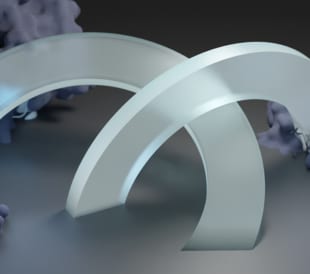
European Molecular Biology Laboratory a Key to Continued Tundra Cryo-TEM Innovation
Cryo-electron microscopy thrives at the European Molecular B...
Read More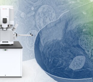
Thermo Fisher Scientific Announces New Plasma-FIB for Volume Electron Microscopy, Cryo-Electron Tomography
Biomedical research leverages volume electron microscopy tec...
Read More
Leave a Reply