Search

Materials Science
Oil and Gas Research
Petroleum science solutions combining electron microscopy and spectroscopy techniques for oil and gas testing.
bf5a- Apps/Techniques/Products/Resources/Contact Us
As demand for oil and gas increases and reserves are depleted, efficient and effective extraction of hydrocarbons is more important than ever. From routine core plug screening to advanced multi-phase flow modeling, Thermo Fisher Scientific provides accurate, end-to-end solutions for oil and gas research and characterization. Whether you are starting a new digital rock analysis lab or expanding your existing characterization capabilities, we have the equipment, software, and expertise you need to maximize your potential from the start.
Core plug screening
A core analysis program is a substantial investment, and assessing the quality of core plugs before special core analysis and geomechanical testing ensures reservoir engineers, petrophysicists, and geologists obtain accurate and representative data before proceeding with further analysis.
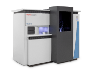
High-resolution imaging offers a non-invasive method to inspect the internal structure of a sample. Thermo Scientific HeliScan microCT (micro-computed tomography) generates a 3D reconstruction of the core plug through one continuous helical X-ray scan. This provides a higher fidelity image than traditional microCT scans due to sophisticated reconstruction algorithms that reduce noise and amplify the signal. These core plug 3D images help petrophysicists and geologists perform multi-scale rock classification for improved understanding of stratigraphy, net to gross, fluid flow, and wireline log response.
- Quickly evaluate sample heterogeneity
- Assess sample contamination (e.g. drilling fluid invasion)
- Determine structural integrity (free from damage caused by drilling)
- Identify the presence and extent of microfractures
Reservoir quality analysis
Every operator has thousands of feet of old core stored in warehouses along with numerous cuttings and thin sections. A digital library of these samples, composed of 3D and 2D images, provides geologists a fast and statistically robust way of evaluating a play by examining previously obtained well cores. These digital rock models combine whole-core computed tomography, microCT, optical and scanning electron microscopy (SEM), and DualBeam technology (focused ion beam and SEM) data through image analysis and visualization software. Previously, these individual observations would be orphaned as traditional methods provide results without context and insight. Digital rock modeling offers analytical and characterization capabilities along with the ability to archive and rapidly share information. Most importantly, it links observed properties to the fundamental nature of the reservoir.
- Obtain accurate clay speciation and textural context with a single method
- Set up automated workflows to guarantee consistent, objective analysis
- Increase confidence in the accuracy of your results

Multi-modal analysis of a core plug with automatic identification of porosity, mineralogy, and oil inclusions.
Multi-scale analysis
Reservoir rocks are dominated by heterogeneity and laminations. To maximize the recovery of hydrocarbons from such reservoirs, accurate characterization of the rock micro-structure is required. This involves not only understanding of the individual rock types and laminations but also the interplay of the various rock types that make up the reservoir. Characterization of subsurface porosity, saturation, and wettability are critical for determining the type and volume of fluids that will be produced. Unfortunately, a single imaging tool cannot resolve both micro-scale pore connectivity and large-scale features.
In order to completely characterize these unconventional systems, multi-scale, multi-modal imaging is required. Thermo Fisher Scientific offers software solutions that correlate X-ray tomography and microscopy imaging with energy-dispersive X-ray spectroscopy (EDS) for elemental analysis. This combination of tools relates microscale observations to the sub-nanometer-scale 3D visualization of pore-system connectivity. It also generates wettability and in situ fluid saturation information, allowing you to upscale microscopic results to the core-plug and log scale.
- Track the distribution of features accurately across scales
- Create statistically valid datasets for representative, quantitative property assessment
- Easily generate automated, objective acquisition and analysis workflows

Multiscale analysis of a core plug from millimeter-scale microCT analysis down to micrometer SEM imaging of two regions of interest.

2D slice from a 3D HeliScan microCT scan of a complex carbonate core plug showing heterogeneity. Voxel size = 10.8 µm.

A composite sample demonstrating the high-contrast imaging of the HeliScan microCT.

Multiscale analysis of a core plug from millimeter-scale microCT analysis down to micrometer SEM imaging of two regions of interest.

Multi-modal analysis of a core plug with automatic identification of porosity, mineralogy, and oil inclusions
2D slice from a 3D HeliScan microCT scan of a complex carbonate core plug showing heterogeneity. Voxel size = 10.8 µm.
A composite sample demonstrating the high-contrast imaging of the HeliScan microCT.
Multiscale analysis of a core plug from millimeter-scale microCT analysis down to micrometer SEM imaging of two regions of interest.
Multi-modal analysis of a core plug with automatic identification of porosity, mineralogy, and oil inclusions

Fundamental Materials Research
Novel materials are investigated at increasingly smaller scales for maximum control of their physical and chemical properties. Electron microscopy provides researchers with key insight into a wide variety of material characteristics at the micro- to nano-scale.
Style Sheet for Komodo Tabs

3D Materials Characterization
Development of materials often requires multi-scale 3D characterization. DualBeam instruments enable serial sectioning of large volumes and subsequent SEM imaging at nanometer scale, which can be processed into high-quality 3D reconstructions of the sample.

Energy Dispersive Spectroscopy
Energy dispersive spectroscopy (EDS) collects detailed elemental information along with electron microscopy images, providing critical compositional context for EM observations. With EDS, chemical composition can be determined from quick, holistic surface scans down to individual atoms.
_Technique_800x375_144DPI.jpg)
EDS Elemental Analysis
Thermo Scientific Phenom Elemental Mapping Software provides fast and reliable information on the distribution of chemical elements within a sample.
_Technique_800x375_144DPI.jpg)
3D EDS Tomography
Modern materials research is increasingly reliant on nanoscale analysis in three dimensions. 3D characterization, including compositional data for full chemical and structural context, is possible with 3D EM and energy dispersive X-ray spectroscopy.
_Technique_800x375_144DPI.jpg)
Environmental SEM (ESEM)
Environmental SEM allows materials to be imaged in their native state. This is ideally suited for academic and industrial researchers who need to test and analyze samples that are wet, dirty, reactive, outgassing or otherwise not vacuum compatible.

Cross-sectioning
Cross sectioning provides extra insight by revealing sub-surface information. DualBeam instruments feature superior focused ion beam columns for high-quality cross sectioning. With automation, unattended high-throughput processing of samples is possible.

Multi-scale analysis
Novel materials must be analyzed at ever higher resolution while retaining the larger context of the sample. Multi-scale analysis allows for the correlation of various imaging tools and modalities such as X-ray microCT, DualBeam, Laser PFIB, SEM and TEM.
3D Materials Characterization
Development of materials often requires multi-scale 3D characterization. DualBeam instruments enable serial sectioning of large volumes and subsequent SEM imaging at nanometer scale, which can be processed into high-quality 3D reconstructions of the sample.
Energy Dispersive Spectroscopy
Energy dispersive spectroscopy (EDS) collects detailed elemental information along with electron microscopy images, providing critical compositional context for EM observations. With EDS, chemical composition can be determined from quick, holistic surface scans down to individual atoms.
EDS Elemental Analysis
Thermo Scientific Phenom Elemental Mapping Software provides fast and reliable information on the distribution of chemical elements within a sample.
3D EDS Tomography
Modern materials research is increasingly reliant on nanoscale analysis in three dimensions. 3D characterization, including compositional data for full chemical and structural context, is possible with 3D EM and energy dispersive X-ray spectroscopy.
Environmental SEM (ESEM)
Environmental SEM allows materials to be imaged in their native state. This is ideally suited for academic and industrial researchers who need to test and analyze samples that are wet, dirty, reactive, outgassing or otherwise not vacuum compatible.
Cross-sectioning
Cross sectioning provides extra insight by revealing sub-surface information. DualBeam instruments feature superior focused ion beam columns for high-quality cross sectioning. With automation, unattended high-throughput processing of samples is possible.
Multi-scale analysis
Novel materials must be analyzed at ever higher resolution while retaining the larger context of the sample. Multi-scale analysis allows for the correlation of various imaging tools and modalities such as X-ray microCT, DualBeam, Laser PFIB, SEM and TEM.
Style Sheet for Instrument Cards Original


Style Sheet to change H2 style to p with em-h2-header class
Style Sheet for Support and Service footer
Style Sheet for Fonts
Style Sheet for Cards
Electron microscopy services for
the materials science
To ensure optimal system performance, we provide you access to a world-class network of field service experts, technical support, and certified spare parts.