Search


Citations & References (66)
Gibco™
Bac-to-Bac™ Baculovirus Expression System
The Bac-to-Bac baculovirus expression system enables the efficient production of recombinant baculovirus for expression testing in insect cells. The systemRead more
| Catalog Number | Quantity |
|---|---|
| 10359016 | 1 kit |
Catalog number 10359016
Price (MXN)
44,218.40
Each
Quantity:
1 kit
Price (MXN)
44,218.40
Each
The Bac-to-Bac baculovirus expression system enables the efficient production of recombinant baculovirus for expression testing in insect cells. The system relies on generation of recombinant baculovirus by site-specific transposition in E. coli rather than homologous recombination in insect cells. This system features:
• Time-saving expression bacmid—With the Bac-to-Bac system, the expression cassette of the pFastBac vector recombines with the parent bacmid in DH10Bac E. coli Competent Cells to form an expression bacmid. The bacmid is then transfected into insect cells for production of recombinant baculovirus particles.
• Easy colony screening—The parent bacmid in DH10Bac E. coli contains a segment of the lacZa gene. The lacZa gene is disrupted upon transposition of the expression cassette into the bacmid, allowing for blue/white selection of recombinants for easier identification of recombinant colonies.
• High transfection efficiency with ExpiFectamine Sf Transfection Reagent—The Bac-to-Bac TOPO expression kits now come with the next-generation ExpiFectamine Sf Transfection Reagent for efficient DNA transfection in insect cells using fast, flexible protocols. Find out more about this reagent ›
• Flexibility—The Bac-to-Bac system comes with a pFastBac vector that contains a large multiple cloning site (MCS) for simplified cloning. Two additional vector formats are offered separately: pFastBac HT vector for production of histidine-tagged recombinant proteins and pFastBac Dual vector for expression of two proteins simultaneously using the p10 and polyhedrin promoters.
• High protein expression—The pFastBac vector uses the strong polyhedrin promoter to generate high levels of expression in a variety of insect cell line such as Sf9, Sf21, and High Five cells.
For Research Use Only. Not for use in diagnostic procedures.
Specifications
Product TypeBaculovirus Expression System
Quantity1 kit
VectorpFastBac
Cloning MethodRestriction Enzyme/MCS
Product LineBac-to-Bac
PromoterPolyhedrin
Protein TagUntagged
Unit SizeEach
Contents & Storage
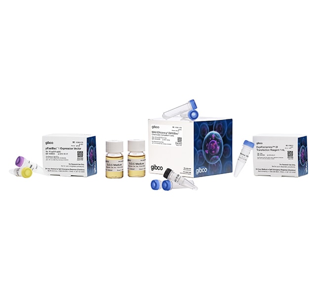
Each Bac-to-Bac Baculovirus Expression System includes:
• pFastBac1 vector (10 μg), store at -20°C
• pFastBac 1-Gus control vector (4 ng), store at -20°C
• MAX Efficiency DH10Bac Chemically Competent E. coli (1 kit with 5 x 0.1 mL), store at -80°C
• ExpiFectamine Sf Transfection Reagent (1 mL), store at 4°C, do not freeze
• pFastBac1 vector (10 μg), store at -20°C
• pFastBac 1-Gus control vector (4 ng), store at -20°C
• MAX Efficiency DH10Bac Chemically Competent E. coli (1 kit with 5 x 0.1 mL), store at -80°C
• ExpiFectamine Sf Transfection Reagent (1 mL), store at 4°C, do not freeze
Frequently asked questions (FAQs)
I cannot grow this white colony in liquid culture. What should I do?
What has happened when I see blue colonies? How about colonies which are blue in the center and white on the edges?
I'm getting mostly white/wild-type plaques instead of blue/recombinant plaques. What am I doing wrong?
I've infected my cells and see large polyhedra in one cell and smaller polyhedra (more numerous) in a neighboring cell. Is this normal?
I'm worried that I am not getting plaques. How many days does it take to see plaques and what size are they typically?
Citations & References (66)
Citations & References
Abstract
The vomeronasal receptor V2R2 does not require escort molecules for expression in heterologous systems.
Journal:Chem Senses
PubMed ID:15647459
'In rodents, many behavioural responses are triggered by pheromones. These molecules are believed to bind and activate two families of G-protein coupled receptors, namely V1Rs and V2Rs, which are specifically expressed in the chemosensory neurons of the vomeronasal organ. V2Rs are homologous with Group 3 of G-protein-coupled receptors, which includes
Identification of Novel SH3 Domain Ligands for the Src Family Kinase Hck. WISKOTT-ALDRICH SYNDROME PROTEIN (WASP), WASP-INTERACTING PROTEIN (WIP), AND ELMO1.
Journal:J Biol Chem
PubMed ID:12029088
'The importance of the SH3 domain of Hck in kinase regulation, substrate phosphorylation, and ligand binding has been established. However, few in vivo ligands are known for the SH3 domain of Hck. In this study, we used mass spectrometry to identify approximately 25 potential binding partners for the SH3 domain
Allocation of helper T-cell epitope immunodominance according to three-dimensional structure in the human immunodeficiency virus type I envelope glycoprotein gp120.
Journal:J Biol Chem
PubMed ID:11551929
'The specificity and intensity of CD4(+) helper T-cell responses determine the effectiveness of immune effector functions. Promiscuously immunodominant helper T-cell epitopes in the human immunodeficiency virus (HIV) envelope glycoprotein gp120 could be important in the development of broadly protective immunity, but the underlying mechanisms of immunodominance and promiscuity remain poorly
Expression, purification, and characterization of an activated cytokine-suppressive anti-inflammatory drug-binding protein 2 (CSBP2) kinase from baculovirus-infected insect cells.
Journal:Protein Expr Purif
PubMed ID:9226723
'An activated form of the human cytokine-suppressive anti-inflammatory drug-binding protein 2 (CSBP2) kinase was expressed in Spodoptera frugiperda (SF9) cells from a baculovirus vector. To maximize expression and to facilitate purification of the recombinant protein, CSBP2 kinase was expressed as a carboxy-terminal fusion protein to glutathione S-transferase (GST). Under optimal
Kinesin Superfamily Motor Protein KIF17 and mLin-10 in NMDA Receptor-Containing Vesicle Transport
Journal:Science
PubMed ID:10846156
'Experiments with vesicles containing N-methyl-D-aspartate (NMDA)receptor 2B (NR2B subunit) show that they are transported alongmicrotubules by KIF17, a neuron-specific molecular motor in neuronaldendrites. Selective transport is accomplished by direct interaction of theKIF17 tail with a PDZ domain of mLin-10 (Mint1/X11), which is aconstituent of a large protein complex including mLin-2