Search

Citations & References (6)
Invitrogen™
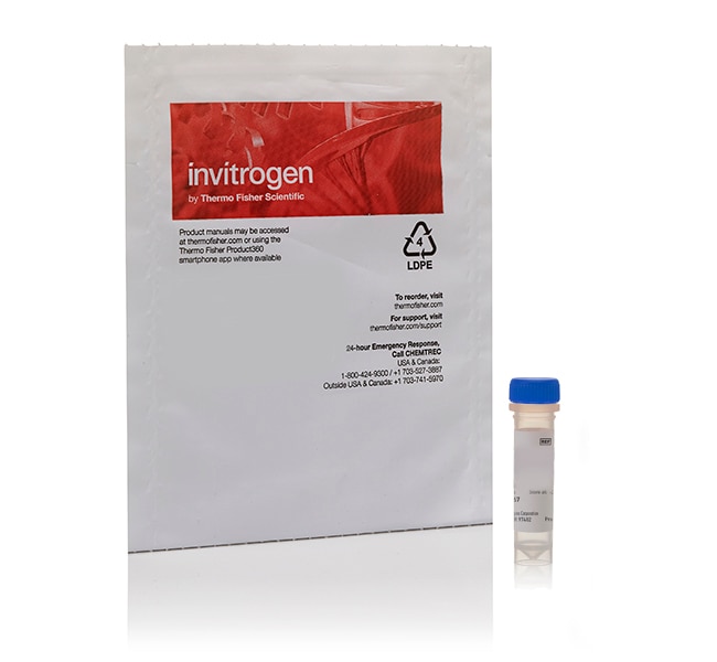
CellLight™ Talin-GFP, BacMam 2.0
CellLight™ Talin-GFP, BacMam 2.0, provides an easy way to label talin with green fluorescent protein (GFP) in live cells. SimplyRead more
| Catalog Number | Quantity |
|---|---|
| C10611 | 1 mL |
Catalog number C10611
Price (USD)
676.00
Each
Quantity:
1 mL
Price (USD)
676.00
Each
CellLight™ Talin-GFP, BacMam 2.0, provides an easy way to label talin with green fluorescent protein (GFP) in live cells. Simply add the reagent to your cells, incubate overnight, and the cells are ready to image in the morning.
Want to label other cell structures? Learn more about CellLight™ fluorescent protein labeling tools
This ready-to-use construct is transduced/transfected into cells using BacMam 2.0 technology, where it expresses GFP fused to the c terminus of human talin. You can observe talin-GFP behavior in live cells to study the focal adhesion of integrins and their interaction with actin filaments using multiple tracking or tracing dyes to image dynamic cellular processes.
Cells expressing CellLight™ constructs can also be fixed with formaldehyde for multiplexed imaging using immunocytochemical techniques.
CellLight™ Technology is:
• Fast and convenient: simply add CellLight™ reagent to your cells, incubate overnight, and image—or store frozen, assay-ready cells for later use
• Highly efficient: up to 90% transduction of a wide range of mammalian cell lines, including primary cells, stem cells, and neurons
• Flexible: co-transduce more than one BacMam reagent for multiplex experiments or co-localization studies; tightly control expression levels by simply varying the dose
• Less toxic: CellLight™ reagents are non-replicating in mammalian cells and are suitable for biosafety level (BSL) 1 handling
BacMam Technology
CellLight™ Talin-GFP, BacMam 2.0, is a fusion construct of the c terminus of human talin and emGFP, providing accurate and specific targeting to cellular talin-GFP. This fusion construct is packaged in the insect virus baculovirus, which does not replicate in human cells and is designated as safe to use with biosafety level (BSL) 1 in most laboratories. BacMam technology ensures that most mammalian cell types are transduced/transfected with high efficiency and minimal toxicity. This transient transfection can be detected after overnight incubation for up to five days—enough time to carry out most dynamic cellular analyses. Like any transfection/transduction technique, the BacMam method does not transfect/transduce all of the cells with equal efficiency, making it poorly suited to cellular population studies or automated imaging/counting. CellLight™ reagents are ideal for experiments where cellular or subcellular co-locatization is required, or for cellular function studies that need special resolution.
For Research Use Only. Not for use in diagnostic procedures.
Specifications
ColorGreen
Detection MethodFluorescence
Dye TypeGFP (EmGFP)
EmissionVisible
Excitation Wavelength Range488⁄510
For Use With (Equipment)Confocal Microscope, Fluorescence Microscope
FormLiquid
Product LineCellLight
Quantity1 mL
Shipping ConditionWet Ice
TechniqueFluorescence Intensity
Label TypeFluorescent Dye
Product TypeTalin
SubCellular LocalizationCytoskeleton
Unit SizeEach
Contents & Storage
Store at 2°C to 6°C, protected from light. Do Not Freeze.
Frequently asked questions (FAQs)
How can I increase the transduction efficiency with the BacMam 2.0 reagents such as the the CellLight and Premo products?
Is there any way to preserve the CellLights labeling beyond 5 days?
Are the CellLights products toxic to cells?
For how long will the CellLights products label my cells?
What cell types can the CellLights products be used with?
Citations & References (6)
Citations & References
Abstract
Baculovirus-mediated gene transfer into mammalian cells.
Journal:Proc Natl Acad Sci U S A
PubMed ID:8637876
This paper describes the use of the baculovirus Autographa californica multiple nuclear polyhedrosis virus (AcMNPV) as a vector for gene delivery into mammalian cells. A modified AcMNPV virus was prepared that carried the Escherichia coli lacZ reporter gene under control of the Rous sarcoma virus promoter and mammalian RNA processing
BacMam technology and its application to drug discovery.
Journal:Expert Opin Drug Discov
PubMed ID:23488908
The recombinant baculovirus/insect cell system was firmly established as a leading method for recombinant protein production when a new potential use for these viruses was revealed in 1995. It was reported that engineered recombinant baculoviruses could deliver functional expression cassettes to mammalian cell types; a system which has come to
Transferrin-targeted porous silicon nanoparticles reduce glioblastoma cell migration across tight extracellular space.
Journal:Sci Rep
PubMed ID:32047170
Stress fiber anisotropy contributes to force-mode dependent chromatin stretching and gene upregulation in living cells.
Journal:Nat Commun
PubMed ID:32994402
Force-activatable coating enables high-resolution cellular force imaging directly on regular cell culture surfaces.
Journal:Phys Biol
PubMed ID:29785968