Search


Citas y referencias (11)
Invitrogen™
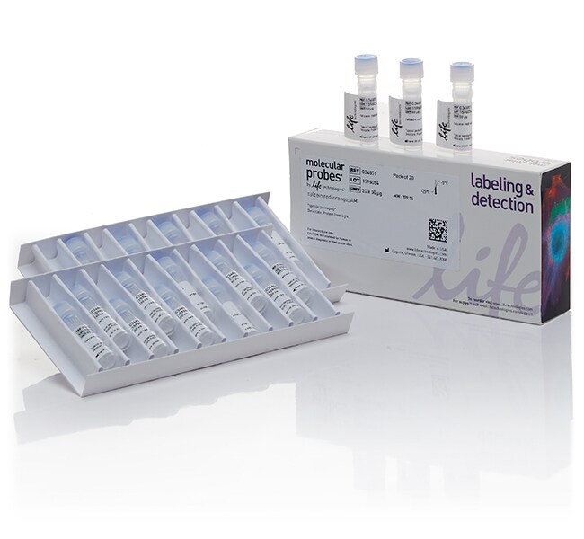
CellTrace™ Calcein Red-Orange, AM - Special Packaging
Calceína AM de color rojo-anaranjado CellTrace™, AM; embalaje especial
| Número de catálogo | Cantidad |
|---|---|
| C34851 | 20 x 50 μg |
Número de catálogo C34851
Precio (MXN)
-
Cantidad:
20 x 50 μg
La calceína AM de color rojo-anaranjado CellTrace es un colorante que penetra en la célula y que puede utilizarse para determinar la viabilidad celular en la mayoría de las células eucariotas. A diferencia de la calceína AM (C-1430, C-3099, C-3100), la calceína AM de color rojo-anaranjado CellTrace es intrínsecamente fluorescente; por lo tanto, se puede necesitar un paso de lavado adicional para minimizar la fluorescencia de fondo del colorante que las células no captan. Sin embargo, la calceína de color rojo-anaranjado CellTrace (excitación/emisión máxima 577/590 nm) se conserva bien en las células vivas que poseen membranas plasmáticas intactas y, por lo tanto, es un marcador celular e indicador de viabilidad celular útil.
Para uso exclusivo en investigación. No apto para uso en procedimientos diagnósticos.
Especificaciones
Permeabilidad celularPermeabilidad celular
DescripciónCalceína AM de color rojo-anaranjado, AM; embalaje especial
Tipo de coloranteOtras etiquetas o colorantes
FormularioLiofilizado
Cantidad20 x 50 μg
Tipo de reactivoCompuestos de seguimiento celular, reactivos de etiquetado celular
Condiciones de envíoTemperatura ambiente
Enzima dianaEsterasa
Emission590 nm
Excitation Wavelength Range577 nm
Para utilizar con (aplicación)Rastreo celular, Rastreador celular
Para utilizar con (equipo)Microscopio de fluorescencia
Línea de productosCellTrace
Tipo de productoTinte
Unit SizeEach
Contenido y almacenamiento
Almacenar en el congelador de -5 °C a -30 °C y proteger de la luz.
Preguntas frecuentes
I need a general cytoplasmic stain that does not overlap with the GFP in my cells. What do you recommend?
How do I reconstitute CellTrace Calcein Red-Orange, AM (Cat. No. C34851)?
I stained two populations of cells, one with CellTracker Green and the other with CellTracker Red, but it looks like there may be crossover of the red dye to the green cells. What is going on?
I stained my cells with Calcein, AM, but the signal went away after I fixed my cells. Why is this?
I'm trying to stain my cells with CellTracker dyes or CFDA SE, but I'm not seeing much signal. What can I do?
Citations & References (11)
Citations & References
Abstract
T lymphocytes expressing a CD16 signaling receptor exert antibody-dependent cancer cell killing.
Journal:
PubMed ID:24197131
To expand applications for T-cell-based immunotherapy in cancer, we designed a receptor that binds the Fc portion of human immunoglobulins and delivers activation signals. The construct included the high-affinity CD16 (FCGR3A) V158 variant, CD8a hinge, and transmembrane domains, along with signaling domains from CD3? and 4-1BB (TNFRSF9), forming a chimeric
H2O2-induced endothelial NO production contributes to vascular cell apoptosis and increased permeability in rat venules.
Journal:Am J Physiol Heart Circ Physiol
PubMed ID:23086988
Although elevated levels of H(2)O(2) have been implicated to play important roles in the pathogenesis of various cardiovascular diseases, the underlying mechanisms remain unclear. This study aims to examine the effect of H(2)O(2) on endothelial nitric oxide (NO) production in intact venules, and elucidate the role and mechanisms of NO
Programmed reduction of ABC transporter activity in sea urchin germline progenitors.
Journal:Development
PubMed ID:22274698
ATP-binding cassette (ABC) transporters protect embryos and stem cells from mutagens and pump morphogens that control cell fate and migration. In this study, we measured dynamics of ABC transporter activity during formation of sea urchin embryonic cells necessary for the production of gametes, termed the small micromeres. Unexpectedly, we found
Connexin-43 upregulation in micrometastases and tumor vasculature and its role in tumor cell attachment to pulmonary endothelium.
Journal:BMC Med
PubMed ID:18647409
The modulation of gap junctional communication between tumor cells and between tumor and vascular endothelial cells during tumorigenesis and metastasis is complex. The notion of a role for loss of gap junctional intercellular communication in tumorigenesis and metastasis has been controversial. While some of the stages of tumorigenesis and metastasis,
The cellular mechanisms of neuronal swelling underlying cytotoxic edema.
Journal:
PubMed ID:25910210
Cytotoxic brain edema triggered by neuronal swelling is the chief cause of mortality following brain trauma and cerebral infarct. Using fluorescence lifetime imaging to analyze contributions of intracellular ionic changes in brain slices, we find that intense Na(+) entry triggers a secondary increase in intracellular Cl(-) that is required for