Search


인용 및 참조 문헌 (7)
Invitrogen™
HCS NuclearMask™ Stains
Easily identify cell nuclei and other features of interest during high-content screening (HCS) assays with the versatile HCS NuclearMask Blue, Red, and Deep Red stains, which can be used with live and fixed cells and in multiparametric analyses.
Have Questions?
보기 방식 변경

| 카탈로그 번호 | 색상 |
|---|---|
| H10325 | Blue |
| H10326 | Red |
| H10294 | Far-Red, Deep Red |
카탈로그 번호 H10325
제품 가격(KRW)
528,000
Each
색상:
Blue
제품 가격(KRW)
528,000
Each
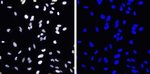
Easily identify cell nuclei in high-content screening (HCS) assays with the versatile HCS NuclearMask Blue, Red, and Deep Red stains, which survive standard formaldehyde-based fixation and detergent-based permeabilization methods and can be applied to live and fixed cells. Because their dyes are easily discriminated from other fluorophores, the HCS NuclearMask stains can be used in multiparametric analyses. They can also label the entire cell (i.e., cytoplasm and nucleus) and provide an accurate backdrop against which to assess the features of interest.
The versatile HCS NuclearMask Blue, HCS NuclearMask Red, and HCS NuclearMask Deep Red stains can be used to measure DNA content and perform cell demarcation with formaldehyde-fixed cells on high content imaging and analysis (HCS) platforms. HCS NuclearMask Blue, HCS NuclearMask Red, and HCS NuclarMask Deep Red stains have excitation/emission maxima of 355/460 nm, 622/645 nm, and 638/686 nm, respectively, when bound to DNA. HCS NuclearMask Blue stain is easily discriminated from visible- and near-IR wavelength fluorophores, HCS NuclearMask Red stain is easily discriminated from green- and even orange-fluorescent fluorophores, and HCS NuclearMask Deep Red is easily discriminated from visible wavelength fluorophores when used in multiparametric analyses.
If total cell staining is desired, HCS CellMask stains label the entire cell (cytoplasm and nucleus) and provide an accurate backdrop against which the features of interest can be assessed. HCS CellMask stains are available in a choice of colors for multiplexing flexibility. Sufficient volume quantities are provided to stain ten 96-well plates using an assay volume of 100 μL per well.
If total cell staining is desired, HCS CellMask stains label the entire cell (cytoplasm and nucleus) and provide an accurate backdrop against which the features of interest can be assessed. HCS CellMask stains are available in a choice of colors for multiplexing flexibility. Sufficient volume quantities are provided to stain ten 96-well plates using an assay volume of 100 μL per well.
For Research Use Only. Not for human or animal therapeutic or diagnostic use.
사양
색상Blue
설명HCS NuclearMask™ Blue Stain
검출 방법Fluorescence
방출Visible
여기 파장 범위350/451
용도(장비)High Content Instrument
제품라인NuclearMask
수량65 μL
배송 조건Room Temperature
라벨 유형Fluorescent Dye
제품 유형Stain
Sub Cellular LocalizationNucleic Acids, Nucleus
Unit SizeEach
구성 및 보관
Includes 65 μL of 2000X concentrate, providing sufficient material to stain a total of ten 96-well microplates.
Store at 2–6°C and protect from light.
Store at 2–6°C and protect from light.
인용 및 참조 문헌 (7)
인용 및 참조 문헌
Abstract
Mammalian cell growth dynamics in mitosis.
Journal:Elife
PubMed ID:31063131
The transcription factor Xrp1 orchestrates both reduced translation and cell competition upon defective ribosome assembly or function.
Journal:Elife
PubMed ID:35179490
Subcellular mRNA Localization Regulates Ribosome Biogenesis in Migrating Cells.
Journal:Dev Cell
PubMed ID:33171110
Pathway-Enriched Gene Signature Associated with 53BP1 Response to PARP Inhibition in Triple-Negative Breast Cancer.
Journal:Mol Cancer Ther
PubMed ID:28958991
Monitoring and modeling of lymphocytic leukemia cell bioenergetics reveals decreased ATP synthesis during cell division.
Journal:Nat Commun
PubMed ID:33020492