Search





Citations & References (20)
Invitrogen™
pHrodo™ Red and Green Dextran, 10,000 MW, for Endocytosis
Perform fast and accurate live-cell endocytosis and imaging assays with pHrodo Red and Green dextran conjugates, which offer pH-sensitive pHrodo dyes that have no need for washing steps or quencher dye, and which can be multiplexed in imaging, HTS, HCA, tracing, and flow applications.
Have Questions?
Change view

| Catalog Number | Color |
|---|---|
| P10361 | Red |
| P35368 | Green |
Catalog number P10361
Price (USD)
572.65
Online exclusive
596.00Save 23.35
Each
Color:
Red
Price (USD)
572.65
Online exclusive
596.00Save 23.35
Each
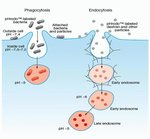
Perform live-cell imaging of endocytosis faster and more accurately without the need for wash steps or quencher dye with pHrodo Red and Green dextran 10,000 MW conjugates. The pH-sensitive pHrodo Red and Green dextran conjugates are nonfluorescent in neutral environments but elicit bright red or green fluorescent signal in pH ranges 5–8, such as those in endosomes and lysosomes. These pHrodo-labeled dextrans can be multiplexed with other dyes and used in imaging, HTS, tracing, and flow applications.
pHrodo Red Dextran (Cat. No. P10361) is a superior alternative to other fluorescent dextran conjugates (e.g., BCECF and tetramethylrhodamine [TRITC]) for live-cell imaging of endocytosis, the process whereby the plasma membrane buds to form membrane-bound vesicles (endosomes), which are then trafficked to various destinations within the cell. pHrodo Red Dextran has pH-sensitive fluorescence excitation/emission spectra of 560/585 nm, and its fluorescence emission increases in intensity with increasing acidity. This increase is particularly dramatic in the range pH 5–8, as commonly seen within endocytic vesicles. pHrodo Red Dextran is essentially dark in the extracellular environment; however, upon internalization, the dextran conjugate elicits a bright, red-fluorescent signal in the acidic environment of the endosomes. pHrodo Red Dextran can also be multiplexed with a wide variety of blue, green, and far-red dyes and reporters such as GFP, Fuo-4, calcein, NucBlue, CellEvent Caspase 3/7 green, Mitosox Green, and Mitotracker Deep Red, among many others.
pHrodo Green Dextran (Cat. No. P35368), like pHrodo Red Dextran, offers fast and accurate results for live-cell endocytosis and phagocytosis assays. pHrodo Green conjugates are non-fluorescent outside the cell at neutral pH, but fluoresce bright green at acidic pH ranges, such as those in endosomes and lysosomes. The pHrodo Green dye can be multiplexed with a wide variety of blue, red, and far-red dye reporters such as Mitosox Red, CellEvent Caspase 3/7 Red, NucBlue, RFPs, and Mitotracker Deep Red, among many others. pHrodo Green Dextran 10,000 MW conjugate can be used in cell imaging, high content screening, high throughput screening, and flow applications. For a broader selection of compatible dyes, please refer to the Cell Staining Tool or SpectraViewer.
Key applications using labeled dextrans:
There are many citations describing the use of labeled dextrans. Some of the most common uses include the following:
• Neuronal tracing (anterograde and retrograde) in live cells
• Cell lineage tracing in live cells
• Neuroanatomical tracing
• Examining intercellular communications (e.g., in gap junctions, during wound healing, and during embryonic development)
• Investigating vascular permeability and blood-brain barrier integrity
• Tracking endocytosis
• Monitoring acidification (some dextran-dye conjugates are pH-sensitive)
• Studying the hydrodynamic properties of the cytoplasmic matrix.
High manufacturing standards
We offer more than 50 fluorescent and biotinylated dextran conjugates in several molecular weight ranges. Dextrans are hydrophilic polysaccharides characterized by their moderate-to-high molecular weight, good water solubility, and low toxicity. They also generally exhibit low immunogeniticy. Dextrans are biologically inert due to their uncommon poly-(α-D-1,6-glucose) linkages, which render them resistant to cleavage by most endogenous cellular glycosidases.
In most cases, our fluorescent dextrans are much brighter and have higher negative charge than dextrans available from other sources. Furthermore, we use rigorous methods for removing as much unconjugated dye as practical, and then assay our dextran conjugates by thin-layer chromatography to help ensure the absence of low molecular weight contaminants.
Wide selection of substituents and molecular weights
Our dextrans are conjugated to biotin or a wide variety of fluorophores, including seven of our Alexa Fluor dyes and are available in these nominal molecular weights (MW): 3,000; 10,000; 40,000; 70,000; 500,000; and 2,000,000 daltons.
pHrodo Green Dextran (Cat. No. P35368), like pHrodo Red Dextran, offers fast and accurate results for live-cell endocytosis and phagocytosis assays. pHrodo Green conjugates are non-fluorescent outside the cell at neutral pH, but fluoresce bright green at acidic pH ranges, such as those in endosomes and lysosomes. The pHrodo Green dye can be multiplexed with a wide variety of blue, red, and far-red dye reporters such as Mitosox Red, CellEvent Caspase 3/7 Red, NucBlue, RFPs, and Mitotracker Deep Red, among many others. pHrodo Green Dextran 10,000 MW conjugate can be used in cell imaging, high content screening, high throughput screening, and flow applications. For a broader selection of compatible dyes, please refer to the Cell Staining Tool or SpectraViewer.
Key applications using labeled dextrans:
There are many citations describing the use of labeled dextrans. Some of the most common uses include the following:
• Neuronal tracing (anterograde and retrograde) in live cells
• Cell lineage tracing in live cells
• Neuroanatomical tracing
• Examining intercellular communications (e.g., in gap junctions, during wound healing, and during embryonic development)
• Investigating vascular permeability and blood-brain barrier integrity
• Tracking endocytosis
• Monitoring acidification (some dextran-dye conjugates are pH-sensitive)
• Studying the hydrodynamic properties of the cytoplasmic matrix.
High manufacturing standards
We offer more than 50 fluorescent and biotinylated dextran conjugates in several molecular weight ranges. Dextrans are hydrophilic polysaccharides characterized by their moderate-to-high molecular weight, good water solubility, and low toxicity. They also generally exhibit low immunogeniticy. Dextrans are biologically inert due to their uncommon poly-(α-D-1,6-glucose) linkages, which render them resistant to cleavage by most endogenous cellular glycosidases.
In most cases, our fluorescent dextrans are much brighter and have higher negative charge than dextrans available from other sources. Furthermore, we use rigorous methods for removing as much unconjugated dye as practical, and then assay our dextran conjugates by thin-layer chromatography to help ensure the absence of low molecular weight contaminants.
Wide selection of substituents and molecular weights
Our dextrans are conjugated to biotin or a wide variety of fluorophores, including seven of our Alexa Fluor dyes and are available in these nominal molecular weights (MW): 3,000; 10,000; 40,000; 70,000; 500,000; and 2,000,000 daltons.
For Research Use Only. Not for human or animal therapeutic or diagnostic use.
Specifications
Cell TypeMammalian
DescriptionpHrodo™ Red Dextran, 10,000 MW, for Endocytosis
Detection MethodFluorescence
Dye TypeOther Label(s) or Dye(s), pH-Sensitive Dyes
Excitation/Emission565/585
FormLyophilized
IndicatorEndocytosis
Quantity0.5 mg
Shipping ConditionRoom Temperature
SolubilitypH >6
Sub Cellular LocalizationEndosomes, Lysosomes
ColorRed
For Use With (Application)Cell Analysis
For Use With (Equipment)Confocal Microscope, Fluorescence Microscope, Flow Cytometer, High Content Analysis Instrument
Product LinepHrodo
Product TypeSupplement
pH5 to 8
Unit SizeEach
Contents & Storage
Store at –20°C, desiccate, and protect from light.
Frequently asked questions (FAQs)
I labeled my cells with pHrodo Red dextran dye to examine endocytosis of my adherent cells. After labeling with the recommended concentration in PBS, the cells rounded up and dissociated. Is this a problem with this reagent?
What is the optimal working concentration for pHrodo Red and Green Dextran, 10,000 MW, for Endocytosis (Cat. Nos. P10361, P35368)?
Citations & References (20)
Citations & References
Abstract
d-Tocopherol Reduces Lipid Accumulation in Niemann-Pick Type C1 and Wolman Cholesterol Storage Disorders.
Journal:J Biol Chem
PubMed ID:23035117
'Niemann-Pick disease type C (NPC) and Wolman disease are two members of a family of storage disorders caused by mutations of genes encoding lysosomal proteins. Deficiency in function of either the NPC1 or NPC2 protein in NPC disease or lysosomal acid lipase in Wolman disease results in defective cellular cholesterol
Molecular basis for pH-dependent mucosal dehydration in cystic fibrosis airways.
Journal:
PubMed ID:24043776
'The ability to maintain proper airway surface liquid (ASL) volume homeostasis is vital for mucus hydration and clearance, which are essential aspects of the mammalian lung''s innate defense system. In cystic fibrosis (CF), one of the most common life-threatening genetic disorders, ASL dehydration leads to mucus accumulation and chronic infection.
Increased acid stability of the hemagglutinin protein enhances H5N1 influenza virus growth in the upper respiratory tract but is insufficient for transmission in ferrets.
Journal:
PubMed ID:23824818
Influenza virus entry is mediated by the acidic-pH-induced activation of hemagglutinin (HA) protein. Here, we investigated how a decrease in the HA activation pH (an increase in acid stability) influences the properties of highly pathogenic H5N1 influenza virus in mammalian hosts. We generated isogenic A/Vietnam/1203/2004 (H5N1) (VN1203) viruses containing either
Host cell autophagy modulates early stages of adenovirus infections in airway epithelial cells.
Journal:J Virol
PubMed ID:23236070
Human adenoviruses typically cause mild infections in the upper or lower respiratory tract, gastrointestinal tract, or ocular epithelium. However, adenoviruses may be life-threatening in patients with impaired immunity and some serotypes cause epidemic outbreaks. Attachment to host cell receptors activates cell signaling and virus uptake by endocytosis. At present, it
Single-stranded DNA oligonucleotides inhibit TLR3-mediated responses in human monocyte-derived dendritic cells and in vivo in cynomolgus macaques.
Journal:Blood
PubMed ID:22700721
TLR3 is a key receptor for recognition of double-stranded RNA and initiation of immune responses against viral infections. However, hyperactive responses can have adverse effects, such as virus-induced asthma. Strategies to prevent TLR3-mediated pathology are therefore desired. We investigated the effect of single-stranded DNA oligonucleotides (ssDNA-ODNs) on TLR3 activation. Human