Search


Citações e referências (58)
Invitrogen™
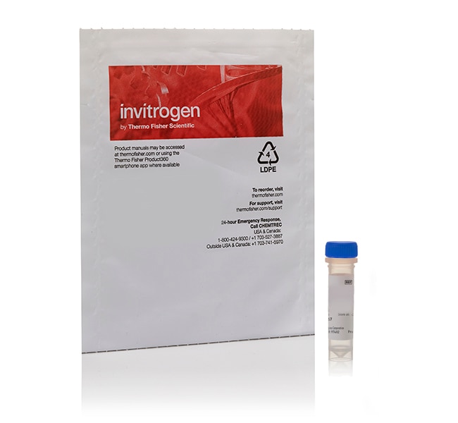
Cholera Toxin Subunit B (Recombinant) Conjugate
Recombinant cholera toxin B subunit (CT-B) conjugates bind with high affinity to GM1 ganglioside and support neuronal tracing, GM1 labeling, membrane trafficking, and membrane organization studies. Choose from Alexa Fluor™ 488, 555, 594, and 647 fluorescent conjugates, Biotin-XX, or horseradish peroxidase (HRP) to match your detection workflow.
Have Questions?
Alterar visualização

| Número do catálogo | Conjugate | Quantity |
|---|---|---|
| C34780 conhecido também como C-34780 | HRP (Horseradish Peroxidase) | 100 μg |
| C22841 | Alexa Fluor 488 | 500 μg |
| C34776 | Alexa Fluor 555 | 100 μg |
| C22843 | Alexa Fluor 555 | 500 μg |
| C34777 | Alexa Fluor 594 | 100 μg |
| C22842 | Alexa Fluor 594 | 500 μg |
| C34778 | Alexa Fluor 647 | 100 μg |
| C34779 | Biotin | 100 μg |
Número do catálogo C34780
conhecido também como C-34780
Preço (BRL)
-
Conjugate:
HRP (Horseradish Peroxidase)
Quantity:
100 μg
Recombinant cholera toxin B conjugates for GM1 labeling and neuronal tracing
Cholera toxin B subunit (CT-B) binds with high affinity to GM1 ganglioside on the cell surface and is widely used for neuronal tracing, GM1 labeling, endocytosis, and membrane trafficking studies. At neutral pH, CT-B forms a pentamer with multiple GM1-binding sites, enabling multivalent interactions with GM1-containing membranes.
These conjugates are made using recombinant cholera toxin B subunit without the enzymatically active A subunit of native cholera toxin.
Why use recombinant cholera toxin B conjugates?
- Label GM1 ganglioside: Use the high-affinity interaction between CT-B and GM1 to visualize and detect GM1-associated membrane structures.
- Trace neuronal projections: Use CT-B for well-established retrograde labeling of neuronal pathways.
- Study membrane trafficking: Follow CT-B/GM1 internalization and retrograde trafficking through endosomal pathways.
- Investigate membrane organization: Label GM1 and examine GM1-associated membrane domains in lipid raft-related studies.
- Use recombinant B subunit: Study GM1 binding and trafficking with CT-B lacking the enzymatically active A subunit of native cholera toxin.
- Choose multiple detection formats: Select fluorescent Alexa Fluor™ conjugates, Biotin-XX, or HRP to match fluorescence, affinity-based, or enzyme-based detection workflows.
Choose a CT-B conjugate for your detection workflow
Select from Alexa Fluor™ 488, 555, 594, and 647 CT-B conjugates for fluorescence imaging, Biotin-XX for avidin- or streptavidin-based detection, or HRP for enzyme-based detection.
Why choose from multiple conjugates?
- Match your imaging system: Select an Alexa Fluor™ conjugate compatible with available excitation sources, emission filters, and detectors.
- Support multicolor imaging: Choose spectrally distinct Alexa Fluor™ dyes for use with other fluorescent probes, antibodies, and cellular markers.
- Enable affinity-based detection: Use Biotin-XX conjugates with avidin- or streptavidin-based detection workflows.
- Enable enzyme-based detection: Use HRP conjugates for chromogenic or chemiluminescent detection workflows.
- Use one GM1-binding probe across workflows: Apply recombinant CT-B with different labels while maintaining the same GM1-binding biological probe.
Applications of cholera toxin B conjugates
Cholera toxin B conjugates are versatile probes for neuronal tracing, GM1 ganglioside labeling, endocytosis, and membrane trafficking studies.
Use CT-B conjugates to:
- Perform neuronal tracing, including widely used retrograde labeling of neuronal projections
- Label GM1 ganglioside on cell membranes
- Visualize GM1-associated membrane domains in lipid raft-related studies
- Study CT-B/GM1 internalization and retrograde trafficking through endosomal pathways
- Perform fluorescence imaging with Alexa Fluor™ 488, 555, 594, or 647 conjugates
- Perform avidin- or streptavidin-based detection with Biotin-XX conjugates
- Perform enzyme-based detection with HRP conjugates
For Research Use Only. Not for use in diagnostic procedures.
For Research Use Only. Not for use in diagnostic procedures.
Especificações
Label TypeOther Label(s) or Dye(s)
Protein FormRecombinant
Protein SubtypeCholera Toxin
Quantity100 μg
Shipping ConditionRoom Temperature
ConjugateHRP (Horseradish Peroxidase)
FormLyophilized
RecombinantRecombinant
Unit SizeEach
Conteúdo e armazenamento
Store in freezer (-5 to -30°C).
Frequently asked questions (FAQs)
I injected a fluorescent tracer, but cannot detect it after tissue is fixed and sectioned. What am I doing wrong?
Do you have a tracer that will only transport retrograde?
How do I know which tracer to choose for my experiment?
What products do you have for neuronal tracing?
Citações e referências (58)
Citações e referências
Abstract
The mechanism of docosahexaenoic acid-induced phospholipase D activation in human lymphocytes involves exclusion of the enzyme from lipid rafts.
Journal:J Biol Chem
PubMed ID:12140281
'Docosahexaenoic acid (DHA), an n-3 polyunsaturated fatty acid that inhibits T lymphocyte activation, has been shown to stimulate phospholipase D (PLD) activity in stimulated human peripheral blood mononuclear cells (PBMC). To elucidate the mechanisms underlying the DHA-induced PLD activation, we first characterized the PLD expression pattern of PBMC. We show
Organization of motoneurons in the dorsal hypoglossal nucleus that innervate the retrusor muscles of the tongue in the rat.
Journal:Anat Rec
PubMed ID:9972807
'This anatomical investigation was prompted by the incomplete knowledge of the myotopic organization of the dorsal subdivison of the hypoglossal nucleus. Intrinsic muscle motoneurons were not segregated and labeled previously with regard to the lateral division of the hypoglossal nerve. Also, motoneuron number and cell size, in relation to the
Peripheral nerve lesion-induced uptake and transport of choleragenoid by capsaicin-sensitive c-fibre spinal ganglion neurons.
Journal:Acta Biol Hung
PubMed ID:12064782
'In the present experiments the effect of systemic capsaicin treatment on the retrograde labelling of sensory ganglion cells was studied following the injection of choleratoxin B subunit-horseradish peroxidase conjugate (CTX-HRP) into intact and chronically transected peripheral nerves. In the control rats CTX-HRP injected into intact sciatic nerves labelled medium and
Localization of amino acids, neuropeptides and cholinergic markers in neurons of the septum-diagonal band complex projecting to the retrosplenial granular cortex of the rat.
Journal:Brain Res Bull
PubMed ID:10974489
'Retrograde labelling was combined with immunohistochemistry to localize neurons containing choline acetyltransferase, gamma-aminobutyric acid (GABA), glutamate, leu-enkephalin, neurotensin, and substance P-like immunoreactivity in the projection pathways from the septum-diagonal band complex to the retrosplenial granular cortex in the rat. Injections of horseradish peroxidase conjugated to subunit B of cholera toxin
Synaptology and ultrastructural characteristics of laryngeal cricothyroid and posterior cricoarytenoid motoneurons in the nucleus ambiguus of the rat.
Journal:Anat Embryol (Berl)
PubMed ID:10463345
'The laryngeal motoneurons innervating the cricothyroid muscle (CT) are located in the semicompact formation just ventral to the rostral part of the compact formation of the nucleus ambiguus. The motoneurons innervating the posterior cricoarytenoid muscle (PCA) are located in the loose formation. We retrogradely labeled the CT and the PCA