Search


Citations & References (3)
Invitrogen™
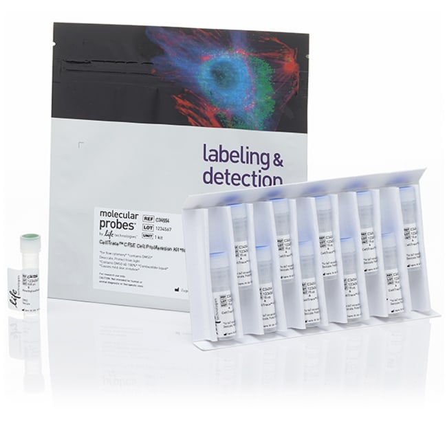
CellTrace™ CFSE Cell Proliferation Kit, for flow cytometry
CellTrace™ CFSE Cell Proliferation Kit is used for in vitro and in vivo labeling of cells to trace multiple generationsRead more
Have Questions?
Change view

| Catalog Number | Sufficient For |
|---|---|
| C34554 | 180 Reactions |
| C34570 | 20 Reactions |
Catalog number C34554
Price (EUR)
292,65
Precio exclusivo compra web
332,00Save 39,35
Each
Sufficient For:
180 Reactions
Price (EUR)
292,65
Precio exclusivo compra web
332,00Save 39,35
Each
CellTrace™ CFSE Cell Proliferation Kit is used for in vitro and in vivo labeling of cells to trace multiple generations using dye dilution by flow cytometry.
• Superior performance—bright, single-peak staining enables visualization of multiple generations
• Long-term signal stability—well-retained in cells for several days post stain
• Non-toxic—staining does not adversely effect cell health
• Simple, robust staining protocol
View a selection guide for all CellTrace™ Cell Proliferation Kits for flow cytometry.
Superior fluorescent staining
Successful proliferation analysis by dye dilution (see figure below) requires an extremely bright dye to distinguish fluorescently labeled cells from auto-fluorescence after several cell divisions. The intense fluorescent staining provided by CellTrace™ CFSE dye enables the visualization of eight or more generations of proliferating cells before the signal is overwhelmed by intrinsic cellular auto-fluorescence. Consistent, homogeneous staining results in very little fluorescence variation between cells in a population, so distinct generations can be seen without any requirement for complex modeling software.
Long-term signal retention
Unlike stains that label the lipid membrane of cells, CellTrace™ CFSE dye easily crosses the plasma membrane and covalently binds inside cells where the stable, well-retained fluorescent dye is designed to provide a consistent signal, even after several days in a cell culture environment.
Non-toxic
CellTrace™ CFSE dye binds covalently to all free amines on the surface and inside of cells and shows little cytotoxicity, with minimal observed effect on the proliferative ability or biology of cells. Researchers have used CFSE SE labeling to show that transplantable hematopoietic cells proliferate in vitro in response to stimulation by a growth factor cocktail.
Simple, robust staining protocol
The CellTrace™ CFSE Cell Proliferation Kit contains convenient single-use vials of dry dye to permit small-scale experiments without preparing excess quantities of dye. A stock solution is prepared by dissolving the contents of a vial in anhydrous DMSO prior to use. To stain 1 mL of cells in protein-free medium, 1 μL of this stock solution is typically used. Cells should be stained for 20 minutes at room temperature with gentle agitation. A brief wash with complete medium will then quench any dye remaining in solution.
Consult user Manual for solubility instructions.
For Research Use Only. Not for use in diagnostic procedures.
Specifications
DescriptionCellTrace™ Cell Proliferation Kit, 180 rxns, CFSE, Lyophilized, Fluorescent Detection Method, 492/517nm Excitation/Emission Wavelength, CellTrace™ CFSE Label/Dye, –5 to –30°C Storage, Use with Flow Cytometer
Detection MethodFluorescence
Dye TypeOther Label(s) or Dye(s)
Emission Wavelength488
FormLyophilized
FormatTube(s)
Quantity180 reactions kit
Reagent TypeCFSE & Related Compounds
Shipping ConditionRoom Temperature
SolubilityDMSO (Dimethylsulfoxide)
Sufficient For180 Reactions
For Use With (Application)Proliferation Assay, Flow Cytometry
For Use With (Equipment)Flow Cytometer
Product LineCellTrace
Product TypeCell Proliferation Kit
Unit SizeEach
Contents & Storage
Contains 10 vials of CellTrace™ CFSE (lyophilized powder) and 1 vial of DMSO (500 μL). Store in freezer (-5°C to -30°C) and protect from light.
Frequently asked questions (FAQs)
I'm trying to label my cells in suspension and track them over time by labeling with CFDA SE, but after labeling them they will no longer adhere to a surface (whereas unlabeled cells adhere well). I've tried to label at 10 and 20 µM. What is causing this?
I want to do a cell migration study for around 4 hours and need to fluorescently label the cells with a dye. What do you recommend?
I labeled my cells with Calcein, AM, but when I imaged the next day, there was no fluorescence from Calcein. Why?
What is the shelf life of CellTrace CFSE Cell Proliferation Kit (Cat. No. C34554, C34570)?
For staining with CellTrace CFSE Cell Proliferation Kit, for flow cytometry (Cat. No. C34554 or C34570), up to how many cells can I have in 1 mL volume?
Citations & References (3)
Citations & References
Abstract
Optimized staining and proliferation modeling methods for cell division monitoring using cell tracking dyes.
Journal:J Vis Exp
PubMed ID:23271219
'Fluorescent cell tracking dyes, in combination with flow and image cytometry, are powerful tools with which to study the interactions and fates of different cell types in vitro and in vivo.(1-5) Although there are literally thousands of publications using such dyes, some of the most commonly encountered cell tracking applications
Appraising the suitability of succinimidyl and lipophilic fluorescent dyes to track proliferation in non-quiescent cells by dye dilution.
Journal:
PubMed ID:25802116
Successful completion of the cell cycle usually results in two identical daughter progeny. This process of generational doubling is termed proliferation and when it occurs in a regulated fashion the benefits range from driving embryonic development to mounting a successful immune response. However when it occurs in a dis-regulated fashion,
TLR4 signaling induced by lipopolysaccharide or paclitaxel regulates tumor survival and chemoresistance in ovarian cancer.
Journal:Oncogene
PubMed ID:19826413
Toll-like receptors (TLRs) expressed on immune cells trigger inflammatory responses. TLRs are also expressed on ovarian cancer (OvCa) cells, but the consequences of signaling by the TLR4/MyD88 pathway in these cells are unclear. Here, TLR4 and MyD88 expression in OvCa tissues (n=20) and cell lines (OVCAR3, SKOV3, AD10, A2780 and