Search


Citations & References (16)
Invitrogen™
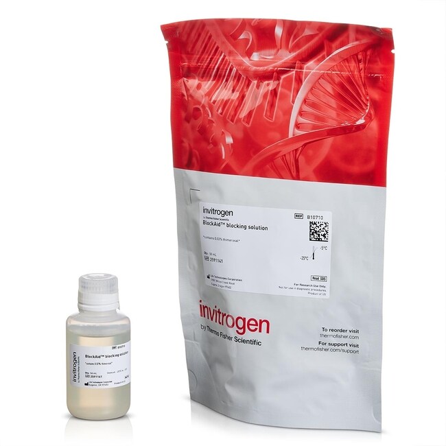
BlockAid™ Blocking Solution
BlockAid™ Blocking Solution is superior to commonly used blocking reagents such as bovine serum albumin, normal serum, or casein forRead more
| Catalog Number | Quantity |
|---|---|
| B10710 | 50 mL |
Catalog number B10710
Price (EUR)
184,65
線上優惠
246,00Save 61,35 (25%)
Each
Quantity:
50 mL
Price (EUR)
184,65
線上優惠
246,00Save 61,35 (25%)
Each
BlockAid™ Blocking Solution is superior to commonly used blocking reagents such as bovine serum albumin, normal serum, or casein for use with cells or tissue sections. It is an optimized mix of protein-blocking components, with no dilution required. BlockAid™ Blocking Solution can be used as the initial blocking reagent before primary antibody is applied to minimize non-specific antibody binding, and as a diluent for both the primary and secondary antibody. This reagent is appropriate for cell or tissue immunofluorescence applications. It has also been validated for use with streptavidin conjugates and microsphere-protein conjugates.
Important Features of BlockAid™ Blocking Solution:
• Excellent background reduction—superior to conventional blocking solutions
• Ready to use—no dilution or stock preparation required
• Versatile—use with any primary or secondary antibody, streptavidin conjugate, microsphere protein conjugates, or Qdot™ conjugates
Minimizing background from non-specific protein binding of antibodies is essential for improved sensitivity and maximum signal-to-background ratios. This is especially crucial when looking for low-expressing antigens, when using samples with high autofluorescence, or for techniques where signals are inherently dim (such as use of directly-labeled primary antibodies or use of super-resolution imaging).
For Research Use Only. Not for use in diagnostic procedures.
Specifications
Quantity50 mL
Reagent TypeBlocking⁄Background Suppression Reagent
Shipping ConditionWet Ice
Product LineBlockAid
TypeBlocking Solution
Unit SizeEach
Contents & Storage
Store in freezer -5°C to -30°C.
Frequently asked questions (FAQs)
Can BlockAid blocking solution be used as a protein blocking solution for antibody labeling on cell or tissue samples, or does it only work with microspheres?
I used a neuron-specific antibody to label my neurons. How can I reduce non-specific antibody binding?
What should I use to block my cells for flow cytometry analysis?
After labeling with my antibody, I am seeing non-specific background binding in my cells or tissue. What could be the cause?
What concentration of my antibody should I use for cell analysis?
Citations & References (16)
Citations & References
Abstract
Restriction of receptor movement alters cellular response: physical force sensing by EphA2.
Journal:Science
PubMed ID:20223987
'Activation of the EphA2 receptor tyrosine kinase by ephrin-A1 ligands presented on apposed cell surfaces plays important roles in development and exhibits poorly understood functional alterations in cancer. We reconstituted this intermembrane signaling geometry between live EphA2-expressing human breast cancer cells and supported membranes displaying laterally mobile ephrin-A1. Receptor-ligand binding,
Proinflammatory and vasodilator effects of nociceptin/orphanin FQ in the rat mesenteric microcirculation are mediated by histamine.
Journal:Am J Physiol Heart Circ Physiol
PubMed ID:17766480
'Nociceptin/orphanin FQ (N/OFQ) is the endogenous ligand for the N/OFQ peptide receptor (NOP). N/OFQ causes hypotension and vasodilation, and we aimed to determine the role of histamine in inflammatory microvascular responses to N/OFQ. Male Wistar rats (220-300 g, n = 72) were anesthetized with thiopental (30 mg/kg bolus, 40-90 mg
Green- and red-fluorescent nanospheres for the detection of cell surface receptors by flow cytometry.
Journal:J Immunol Methods
PubMed ID:9831388
'Fluorescent probes serve as sensitive tools for obtaining structural and functional information in cellular systems. In spite of the high sensitivity provided by fluorescent reagents, cell surface receptors expressed in low numbers often escape detection with commonly used fluorescent probes. R-Phycoerythrin (R-PE), a molecule with a very high quantum yield,
Micromechanical tests of adhesion dynamics between neutrophils and immobilized ICAM-1.
Journal:Biophys J
PubMed ID:14747356
'Strong, integrin-mediated adhesion of neutrophils to endothelium during inflammation is a dynamic process, requiring a conformational change in the integrin molecule to increase its affinity for its endothelial counterreceptors. To avoid general activation of the cell, Mg(2+) was used to induce the high-affinity integrin conformation, and micromechanical methods were used
Reversible self-assembly and directed assembly of DNA-linked micrometer-sized colloids.
Journal:Proc Natl Acad Sci U S A
PubMed ID:15758072
'We present a technique for the directed assembly and self-assembly of micrometer-scale structures based on the control of specific DNA linkages between colloidal particles. The use of DNA links combined with polymer brushes provides an effective way to regulate the range and magnitude of addressable forces between pairs (and further