Search


Citations & References (3)
Invitrogen™
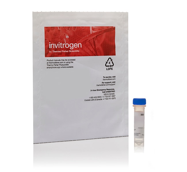
Qdot™ 525 Streptavidin Conjugate
Qdot™ 525 streptavidin conjugate comprises a biotin-binding protein (streptavidin) covalently attached to a fluorescent label (Qdot™ nanocrystal). Streptavidin has aRead more
Have Questions?
Change view

| Catalog Number | Quantity |
|---|---|
| Q10143MP | 50 μL |
| Q10141MP | 200 μL |
Catalog number Q10143MP
Price (INR)
-
Quantity:
50 μL
Qdot™ 525 streptavidin conjugate comprises a biotin-binding protein (streptavidin) covalently attached to a fluorescent label (Qdot™ nanocrystal). Streptavidin has a very high binding affinity for biotin, and a conjugate of streptavidin is commonly used together with a conjugate of biotin for specific detection of a variety of proteins, protein motifs, nucleic acids, and other molecules (for example, a biotinylated primary antibody bound to a protein target can be detected with a fluorescently labeled streptavidin). Strategies similar to this are used in many detection protocols including western blots, flow cytometry, imaging and microscopy, and microplate assays, and are also used in purification workflows to achieve targeted fractionation. Qdot™ nanocrystal conjugates are provided as 1 μM solutions.
Important Features of Qdot™ Streptavidin Conjugates:
• Streptavidin Qdot™ 525 conjugate has emission maxima of ∼525 nm
• Approximately 5 to 10 streptavidins per Qdot™ nanocrystal
• Extremely photostable and bright fluorescence
• Efficiently excited with single-line excitation sources
• Narrow emission, large Stokes shift
• Available in multiple colors
• Ideal for western blots, flow cytometry, imaging and microscopy, microplate assays and more
Properties of Qdot™ Nanocrystals
The Qdot™ streptavidin conjugate is the size of a large macromolecule or protein (∼15–20 nm) and represents the brightest class of streptavidin detection reagents. Qdot™ streptavidin conjugates are made from a nanometer-scale crystal of a semiconductor material (CdSe), which is coated with an additional semiconductor shell (ZnS) to improve the optical properties of the material. The Qdot™ 705 and Qdot™ 800 streptavidin conjugates, which include CdSeTe, are made in a similar fashion. This core-shell material is further coated with a polymer shell that allows the materials to be conjugated to biological molecules and to retain their optical properties.
Other Fluorescent Conjugates of Streptavidin are Available
We offer several other Qdot™ colors or try the Qdot™ Streptavidin Sampler Kit, containing Qdot™ streptavidin conjugates in six colors 525, 565, 585, 605, 655, and 705. In addition to nanocrystal conjugates, we offer a wide range of streptavidins conjugated to Alexa Fluor™ dyes, Oregon Green™ dye, enzyme conjugates, and traditional fluorophores like Texas Red™ dye, fluorescein (FITC) and more.
Find Biotinylated Conjugates
We offer an extensive line of biotinylated conjugates for use in biotin-streptavidin detection strategies.
• Use the primary antibody search tool to find biotinylated primary antibodies
• Use the secondary antibody selector tool to find biotinylated secondary antibodies and biotinylated anti-dye and anti-hapten antibodies
Blocking Endogenous Biotin
Naturally occurring biotins can interfere with biotin-streptavidin detection schemes. For experiments involving fixed and permeabilized cells, try our Endogenous Biotin-Blocking Kit to minimize this interference.
For Research Use Only. Not intended for any animal or human therapeutic or diagnostic use.
Related Links:
Learn more about Avidin-Biotin Detection
To learn more about Qdot™ Nanocrystals
Important Features of Qdot™ Streptavidin Conjugates:
• Streptavidin Qdot™ 525 conjugate has emission maxima of ∼525 nm
• Approximately 5 to 10 streptavidins per Qdot™ nanocrystal
• Extremely photostable and bright fluorescence
• Efficiently excited with single-line excitation sources
• Narrow emission, large Stokes shift
• Available in multiple colors
• Ideal for western blots, flow cytometry, imaging and microscopy, microplate assays and more
Properties of Qdot™ Nanocrystals
The Qdot™ streptavidin conjugate is the size of a large macromolecule or protein (∼15–20 nm) and represents the brightest class of streptavidin detection reagents. Qdot™ streptavidin conjugates are made from a nanometer-scale crystal of a semiconductor material (CdSe), which is coated with an additional semiconductor shell (ZnS) to improve the optical properties of the material. The Qdot™ 705 and Qdot™ 800 streptavidin conjugates, which include CdSeTe, are made in a similar fashion. This core-shell material is further coated with a polymer shell that allows the materials to be conjugated to biological molecules and to retain their optical properties.
Other Fluorescent Conjugates of Streptavidin are Available
We offer several other Qdot™ colors or try the Qdot™ Streptavidin Sampler Kit, containing Qdot™ streptavidin conjugates in six colors 525, 565, 585, 605, 655, and 705. In addition to nanocrystal conjugates, we offer a wide range of streptavidins conjugated to Alexa Fluor™ dyes, Oregon Green™ dye, enzyme conjugates, and traditional fluorophores like Texas Red™ dye, fluorescein (FITC) and more.
Find Biotinylated Conjugates
We offer an extensive line of biotinylated conjugates for use in biotin-streptavidin detection strategies.
• Use the primary antibody search tool to find biotinylated primary antibodies
• Use the secondary antibody selector tool to find biotinylated secondary antibodies and biotinylated anti-dye and anti-hapten antibodies
Blocking Endogenous Biotin
Naturally occurring biotins can interfere with biotin-streptavidin detection schemes. For experiments involving fixed and permeabilized cells, try our Endogenous Biotin-Blocking Kit to minimize this interference.
For Research Use Only. Not intended for any animal or human therapeutic or diagnostic use.
Related Links:
Learn more about Avidin-Biotin Detection
To learn more about Qdot™ Nanocrystals
For Research Use Only. Not for use in diagnostic procedures.
Specifications
Concentration1 μM
Product TypeStreptavidin Conjugate (fluorescent)
Quantity50 μL
Shipping ConditionRoom Temperature
ConjugateQdot 525
FormLiquid
Product LineQdot
Unit SizeEach
Contents & Storage
1 tube — Store at 2 to 6°C; DO NOT FREEZE
Frequently asked questions (FAQs)
I used a neuron-specific antibody to label my neurons. I can't get enough signal from my fluorescent dye conjugated primary antibody. What can I do to improve it?
I have a very low-abundance antigen. How can I amplify my signal?
I am getting very high background with my Qdot streptavidin conjugate. Do you have any suggestions?
I am getting no signal with my Qdot streptavidin conjugate. What should I do?
What is the best way to remove white precipitate from my ITK Qdot nanocrystals?
Citations & References (3)
Citations & References
Abstract
Immunomodulatory effects of HSV-2 infection on immature macaque dendritic cells modify innate and adaptive responses.
Journal:Blood
PubMed ID:15845898
'Herpes simplex viruses (HSV) infect human and murine dendritic cells (DCs) and interfere with their immunostimulatory functions in culture. HSV-2 infection increases human immunodeficiency virus (HIV) spread in patients, and DCs also promote HIV infection. We have studied these topics in rhesus macaque monocyte-derived DCs (moDCs) to set the stage
TDAG51 mediates epithelial-to-mesenchymal transition in human proximal tubular epithelium.
Journal:Am J Physiol Renal Physiol
PubMed ID:22592641
'Epithelial-to-mesenchymal transition (EMT) contributes to renal fibrosis in chronic kidney disease. Endoplasmic reticulum (ER) stress, a feature of many forms of kidney disease, results from the accumulation of misfolded proteins in the ER and leads to the unfolded protein response (UPR). We hypothesized that ER stress mediates EMT in human
Delivery of siRNA and other macromolecules into skin and cells using a peptide enhancer.
Journal:Proc Natl Acad Sci U S A
PubMed ID:21903933
'Delivery of macromolecules into cells and tissues such as skin is a major challenge. This obstacle poses a particular challenge for the delivery of siRNA where cellular and tissue level transport barriers need to be overcome. siRNAs are potential therapeutics for various dermatological diseases including psoriasis, atopic dermatitis, and cancer;