Search


Citations & References (10)
Invitrogen™
Click-iT™ Plus TUNEL Assay Kits for In Situ Apoptosis Detection
Detect apoptosis in cells and tissues samples with Click-iT Plus TUNEL Assay kits, which offer easy dye incorporation and can be multiplexed with GFP and RFP.
Have Questions?
Change view

| Catalog Number | Color | Label or Dye |
|---|---|---|
| C10618 | Red | Alexa Fluor™ 594 |
| C10617 | Green | Alexa Fluor™ 488 |
| C10619 | Far-Red | Alexa Fluor™ 647 |
Catalog number C10618
Price (KRW)
-
Color:
Red
Label or Dye:
Alexa Fluor™ 594
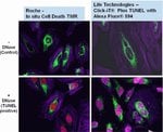
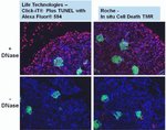
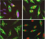
Detect more apoptotic cells in tissues and cultured cell samples with the Click-iT Plus TUNEL Assay for In Situ Apoptosis Detection, which offers Alexa Fluor 488, 594, and 647 fluorescent dye options. This in situ apoptosis detection kit is optimized for tissue or cell samples and the dyes can be multiplexed with other dyes or proteins, such as GFP and RFP, and incorporated more readily into complex molecules due to their smaller size (compared with antibodies). This TUNEL assay kit is also very flexible and can be used to test 1–50 samples in a single experiment.
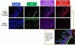
The Click-iT Plus TUNEL Alexa Fluor 488, 594, and 647 assays for in situ apoptosis detection can detect apoptotic cells in tissue and cultured cell samples through the incorporation of a small, highly specific labeling moiety and a bright fluorescent dye. After incorporation of the labeling moiety into DNA fragments, detection is achieved through a catalyzed “click” reaction using conditions mild enough to preserve the emitted fluorescent signal from GFP or RFP.
Other advantages of the Click-iT Plus TUNEL Assay for In Situ Apoptosis Detection include:
• Optimized for the detection of apoptotic cells in either tissue or cell samples
• Multiplex enabled—optimized to work with fluorescent dyes or proteins such as GFP and RFP
• Improved TUNEL assay—better label incorporation due to small reactive moiety
• Bright apoptotic signal—uses Alexa Fluor dyes, resulting in a stable, non-photobleaching fluorescent signal
• Flexibility—the assay can be configured to test 1–50 samples at a time
Fragmentation of cellular DNA is a hallmark of apoptosis. The TUNEL assay is the most widely used method to detect fragmented DNA in apoptotic cells or tissue samples. The TUNEL assay begins with incorporation of modified dUTP at the 3’-OH end of the fragmented DNA. The dUTP modification is often the addition of a fluorophore. Due to the size of the fluorophore, the modified dUTP can display lower than expected incorporation rates, which can affect the sensitivity of the TUNEL assay. Additionally, many fluorophores used in currently available TUNEL assay kits suffer from photobleaching and fluorescent spectral overlap issues, both of which reduce the sensitivity of and ability to multiplex the assay.
The Click-iT Plus TUNEL assay was developed to address these issues. The assay uses dUTP modified with an alkyne group (a small bio-orthogonal functional group), allowing the nucleotide to be more readily incorporated. After incorporation, a highly specific click reaction between the alkyne group and an Alexa Fluor picolyl azide fluorescent dye, and subsequent detection of that dye, results in a sensitive and specific assay for the detection of apoptotic cells or tissue samples. Because of its gentle reaction conditions, the Click-iT Plus TUNEL assay enables multiplexing with fluorescent proteins or dyes.
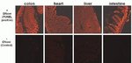
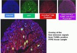
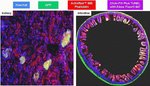
The Click-iT Plus TUNEL assay has been validated with several different formalin-fixed paraffin-embedded tissue types. In all cases, its ability to multiplex with fluorescent proteins and dyes was preserved. Additionally, the ability to stain actin using fluorescent-labeled phalloidin was also preserved.
The Click-iT Plus TUNEL assay contains all the reagents needed to detect apoptotic cells from either tissue or cell samples. The reagents supplied in this kit can be used to test 50 samples and can be configured to test 1–50 samples at a time.
Other advantages of the Click-iT Plus TUNEL Assay for In Situ Apoptosis Detection include:
• Optimized for the detection of apoptotic cells in either tissue or cell samples
• Multiplex enabled—optimized to work with fluorescent dyes or proteins such as GFP and RFP
• Improved TUNEL assay—better label incorporation due to small reactive moiety
• Bright apoptotic signal—uses Alexa Fluor dyes, resulting in a stable, non-photobleaching fluorescent signal
• Flexibility—the assay can be configured to test 1–50 samples at a time
Fragmentation of cellular DNA is a hallmark of apoptosis. The TUNEL assay is the most widely used method to detect fragmented DNA in apoptotic cells or tissue samples. The TUNEL assay begins with incorporation of modified dUTP at the 3’-OH end of the fragmented DNA. The dUTP modification is often the addition of a fluorophore. Due to the size of the fluorophore, the modified dUTP can display lower than expected incorporation rates, which can affect the sensitivity of the TUNEL assay. Additionally, many fluorophores used in currently available TUNEL assay kits suffer from photobleaching and fluorescent spectral overlap issues, both of which reduce the sensitivity of and ability to multiplex the assay.
The Click-iT Plus TUNEL assay was developed to address these issues. The assay uses dUTP modified with an alkyne group (a small bio-orthogonal functional group), allowing the nucleotide to be more readily incorporated. After incorporation, a highly specific click reaction between the alkyne group and an Alexa Fluor picolyl azide fluorescent dye, and subsequent detection of that dye, results in a sensitive and specific assay for the detection of apoptotic cells or tissue samples. Because of its gentle reaction conditions, the Click-iT Plus TUNEL assay enables multiplexing with fluorescent proteins or dyes.
The Click-iT Plus TUNEL assay has been validated with several different formalin-fixed paraffin-embedded tissue types. In all cases, its ability to multiplex with fluorescent proteins and dyes was preserved. Additionally, the ability to stain actin using fluorescent-labeled phalloidin was also preserved.
The Click-iT Plus TUNEL assay contains all the reagents needed to detect apoptotic cells from either tissue or cell samples. The reagents supplied in this kit can be used to test 50 samples and can be configured to test 1–50 samples at a time.
For Research Use Only. Not for use in diagnostic procedures.
Specifications
ColorRed
DescriptionClick-iT Plus TUNEL Assay for In Situ Apoptosis Detection, Alexa Fluor™ 594 dye
Excitation/Emission590/617
For Use With (Equipment)Fluorescence Microscope
Label TypeAlexa Fluor™ Dyes
Label or DyeAlexa Fluor™ 594
No. of Reactions50 coverslips
Product LineClick-iT
Product TypeTUNEL Assay
Quantity1 kit
Shipping ConditionDry Ice
Storage RequirementsStore at ≤20°C and protect from light.
Detection MethodFluorescence
FormatCoverslip
Unit SizeEach
Frequently asked questions (FAQs)
I will be performing a cell proliferation assay using Click-iT EdU kit. At what point can I stop overnight, or do I have to perform all the steps continuously?
A control for a Click-iT EdU labeling experiment uses no EdU and the Click-iT reaction using Alexa Fluor 594 azide. The mouse heart tissue sections are showing non-specific labeling in red, seen in particular clusters of cells. They don't overlap with DAPI. What is the problem?
I need to test cells for apoptosis after they have been formaldehyde-fixed and permeabilized. What dye or conjugate do you recommend? Will Annexin V conjugates work?
Can I use Click-iT TUNEL Alexa Fluor Imaging Assays for Microscopy & HCS (Cat. No. C10246) for flow cytometry?
Can I use Click-iT Plus TUNEL Assay Kits for In Situ Apoptosis Detection (Cat. Nos. C10617, C10618, C10619) for whole mount immunofluorescence staining of zebrafish larvae?
Citations & References (10)
Citations & References
Abstract
CD13 deficiency leads to increased oxidative stress and larger atherosclerotic lesions.
Journal:Atherosclerosis
PubMed ID:31229835
'Atherosclerosis is an inflammatory cardiovascular disorder characterized by accumulation of lipid-loaded macrophages in the intima. Prolonged accumulation leads to apoptosis of macrophages and eventually to progression of lesion development. Prevention of macrophage accumulation within the intima has been shown to reduce lesion formation. Since CD13 mediates trafficking of macrophages to
Loss of host-derived osteopontin creates a glioblastoma-promoting microenvironment.
Journal:Neuro Oncol
PubMed ID:29016864
'Microglia and periphery-derived monocytes infiltrate human and mouse glioblastoma and their density is positively correlated with malignancy. Using microarray and RNA sequencing, we have previously shown that glioblastoma-associated microglia/monocytes (GAMs) express osteopontin/SPP1.'
Differential susceptibility of mouse strains on pancreatic injury and regeneration in cerulein-induced pancreatitis.
Journal:Int J Clin Exp Pathol
PubMed ID:31966883
'Acute pancreatitis (AP), a common disease, causes significant morbidity and mortality in clinical practice. Our objective of this study was to establish an experimental mouse AP model with cerulein treatment and to explore the susceptibility of mouse strains on the severity of pancreatic injury and the subsequent repair and regeneration.
Tanshinone IIA promotes IL2-mediated SW480 colorectal cancer cell apoptosis by triggering INF2-related mitochondrial fission and activating the Mst1-Hippo pathway.
Journal:Biomed Pharmacother
PubMed ID:30372868
IL-2-based therapy is a promising tool to treat colorectal cancer, but drug resistance always occurs in clinical practice. Mitochondrial fission is a novel target to modulate cancer development and progression. The aim of our study is to explore the effect of IL-2 combined with Tan IIA on SW480 colorectal cancer
Sirtuin-1 protects hair follicle stem cells from TNFa-mediated inflammatory stress via activating the MAPK-ERK-Mfn2 pathway.
Journal:Life Sci
PubMed ID:30292830
Stem cell transplantation is a promising tool to treat burn injuries. However, the inflammatory microenvironment in damaged skin limits the efficiency of stem cell-based therapy via poorly understood mechanisms. The aim of our study is to explore the contribution and mechanism of Sirtuin-1 (Sirt1) in TNFa-mediated inflammatory stress in hair