Search


인용 및 참조 문헌 (1)
Invitrogen™
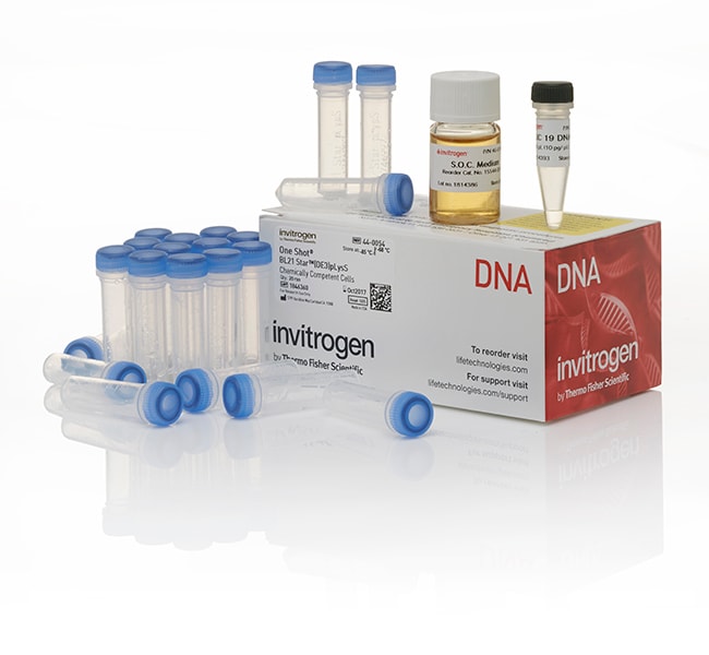
BL21 Star™ (DE3)pLysS One Shot™ Chemically Competent E. coli
One Shot™ BL21 Star™ (DE3) pLysS E. coli는 발현율이 높은 T7 promoter 기반 발현 시스템 (예: pRSET T7 벡터)에서 비독성자세히 알아보기
| 카탈로그 번호 | 수량 |
|---|---|
| C602003 | 21 x 50 μL/tube |
카탈로그 번호 C602003
제품 가격(KRW)
637,000
온라인 행사
Ends: 30-Sep-2026
728,000할인액 91,000 (13%)
Each
수량:
21 x 50 μL/tube
제품 가격(KRW)
637,000
온라인 행사
Ends: 30-Sep-2026
728,000할인액 91,000 (13%)
Each
One Shot™ BL21 Star™ (DE3) pLysS E. coli는 발현율이 높은 T7 promoter 기반 발현 시스템 (예: pRSET T7 벡터)에서 비독성 재조합 단백질의 고발현이 필요한 어플리케이션을 위해 설계된 chemically competent cell입니다. One Shot™ BL21 Star™ (DE3) pLysS Chemically Competent 세포는 transformation efficiency가 >1 x 10⁸ cfu/ μg plasmid DNA입니다.
• 높은 mRNA 안정성으로 단백질 수율이 높습니다.
• 발현율이 높은 T7 promoter 기반 plasmid에 사용하기 이상적입니다.
• 발현이 유도되지 않은 세포에서 background 발현이 낮습니다.
높은 mRNA 안정성으로 단백질 수율 향상
One Shot™ BL21 Star™ (DE3) pLysS E. coli는 mRNA 안정성이 높아 단백질 발현에 충분한 mRNA를 이용할 수 있습니다. 이렇게 안정성이 높은 이유는 mRNA 분해에 관여하는 RNaseE 유전자 돌연변이(rne131) 때문입니다.
비독성 재조합 단백질의 높은 발현
One Shot™ BL21 Star™ (DE3) pLysS 세포는 발현율이 높은 T7 promoter 기반 발현 시스템에서 비독성이지만 잠재적으로 성장을 억제하는 재조합 단백질의 높은 발현에 이상적입니다. BL21 Star™ (DE3) pLysS 균주에 있는 pLysS CamR Plasmid는 유도되지 않은 세포에서 단백질의 발현을 방지하는 T7 RNA 중합효소 억제제인 T7 lysozyme을 발현합니다. pLysS의 p15a origin은 이 plasmid가 pUC 또는 pBR322 유래 plasmid와 호환되게 합니다. 이 균주는 lacUV5 promoter 제어 하에서 T7 RNA 중합효소 유전자를 가진 DE lysogen도 함유하고 있으며 T7 RNA 중합효소 발현을 유도하기 위해 IPTG가 필요합니다. One Shot™ BL21 Star™ (DE3) pLysS 세포에는 lon protease가 들어있지 않으며 외막 protease, OmpT도 없습니다. 이런 protease의 결핍은 이종 단백질의 분해를 줄입니다.
참고: BL21 Star™ strains은 mRNA 안정성이 높아 BL21 균주보다 이종 유전자의 기본 발현이 높습니다. 그러므로 이런 균주는 독성 유전자 발현에 유용하지 않을 수 있습니다. 하지만 BL21 Star™ (DE3) pLysS 균주는 BL21 Star™ 균주보다 발현 수준이 낮습니다.
유전자형:
F-ompT hsdSB (rB-, mB-) galdcmrne131 (DE3) pLysS (CamR)
사용하기 쉬운 Single Tube 형식
일회용 Single Tube 형식으로 플레이트에 옮길 때까지 같은 tube에서 모든 transformation 단계를 실시할 수 있어 시간을 절약하고 오염을 방지할 수 있습니다.
필요한 균주 및 형식 검색
본사는 chemically competent cells 및 electrocompetent cells의 여러 균주와 형식을 제공하여 여러분의 특정 transformation 필요에 부합하도록 합니다. 독성 단백질 발현에는 BL21-AI™ One Shot™ Chemically Competent E. coli를 선택하십시오. 복제수가 적은 T7 promoter 기반 발현 시스템에서 비독성 재조합 단백질의 발현은(예: Champion™ pET vectors) 자사의 One Shot™ BL21 Star™ (DE3) chemically competent cells을 선택하십시오.
연구용으로만 사용할 수 있습니다. 치료 또는 진단 목적으로 동물이나 사람에게 사용할 수 없습니다.
For Research Use Only. Not for use in diagnostic procedures.
사양
항생제 내성 박테리아Yes (Chloramphenicol)
블루/화이트 스크리닝No
메틸화 DNA 클로닝No
에프에피솜 포함No
고처리량 호환성Low
플라스미드 품질 개선No
Improves Protein StabilityYes (lon, ompT)
Improves RNA StabilityYes (rne131, pLysS)
비메틸화 DNA 준비Yes (dcm)
제품라인One Shot
제품 유형Chemically Competent Cells
수량21 x 50 μL/tube
재조합 감소No
배송 조건Dry Ice
T1 Phage - 저항성(tonA)No
Toxic ProteinsNo
형질전환 효율 수준Medium Efficiency (1 x 10⁸ to 1 x 10⁹ cfu/μg)
형식Tube
프로모터T7
종E. coli (B)
Unit SizeEach
구성 및 보관
• BL21 Star (DE3)pLysS Chemically Competent E. coli (21 x 50 μL); storeat –80°C
• pUC19 DNA (1 x 50 μL); store at –20°C
• S.O.C. Medium (6 mL); store at 4°C or room temperature
• pUC19 DNA (1 x 50 μL); store at –20°C
• S.O.C. Medium (6 mL); store at 4°C or room temperature
자주 묻는 질문(FAQ)
My gene of interest is toxic to bacterial cells. Are there any precautions you can suggest?
I'm trying to express my protein using a bacterial expression system. How do I know if I'm seeing degradation of my protein or if what I’m seeing is codon usage bias?
I'm trying to express my protein using a bacterial expression system and am getting inclusion bodies. What should I do?
I'm getting low protein yield from my bacterial expression system. What can I do to improve this?
My cells are growing very slowly, and I'm not getting any protein expression from my baterial expression system. What can I do to fix this?
인용 및 참조 문헌 (1)
인용 및 참조 문헌
Abstract
Mertk triggers uptake of photoreceptor outer segments during phagocytosis by cultured retinal pigment epithelial cells.
Journal:J Biol Chem
PubMed ID:11861639
The RCS rat is a widely studied model of recessively inherited retinal degeneration. The genetic defect, known as rdy (retinal dystrophy), results in failure of the retinal pigment epithelium (RPE) to phagocytize shed photoreceptor outer segment membranes. We previously used positional cloning and in vivo genetic complementation to demonstrate that