SeeBlue™ Plus2 Pre-stained Protein Standard
Invitrogen™

SeeBlue™ Plus2 Pre-stained Protein Standard

SeeBlue™ Plus2 Pre-Stained Standard에는 단백질 10개가 함유되어 있습니다. 이중 8개는 blue-dye되고 2개는 contrasting color로 나타나 밴드 확인이 쉽습니다. SeeBlue™ Plus2자세히 알아보기
Have Questions?
카탈로그 번호수량
LC5925500 μL
카탈로그 번호 LC5925
제품 가격(KRW)
270,000
Online offer
Ends: 31-Dec-2025
300,000
할인액 30,000 (10%)
Each
카트에 추가하기
수량:
500 μL
대량 주문 또는 맞춤형 요청
제품 가격(KRW)
270,000
Online offer
Ends: 31-Dec-2025
300,000
할인액 30,000 (10%)
Each
카트에 추가하기
SeeBlue™ Plus2 Pre-Stained Standard에는 단백질 10개가 함유되어 있습니다. 이중 8개는 blue-dye되고 2개는 contrasting color로 나타나 밴드 확인이 쉽습니다. SeeBlue™ Plus2 Standard는 전기영동 중 분자량을 쉽게 가시화하여 western transfer efficiency를 신속하게 평가할 수 있습니다. NuPAGE™, Tris-Glycine, Tricine Gel과 함께 사용할 수 있습니다. SeeBlue™ Plus2 Standard 특징:

• 10 polypeptides가 4 kDa ∼250 kDa 범위에서(buffer system에 따라) blue 8개, colored band 2개로 분해됩니다.

• 즉각적으로 모호한 밴드 확인

• ready-to-use format으로 mixing, heating, reducing이 필요하지 않습니다.

참고: 이 standard는 pre-reduced되어 있어 loading buffer에 reducing reagent가 함유되지 않습니다.

본 제품은 냉장/냉동제품으로 반송된 제품은 전량 폐기 처리 되오니 주문 전 상세 내용 다시 한번 확인 부탁드립니다.
For Research Use Only. Not for use in diagnostic procedures.
사양
검출 방법Colorimetric
젤 호환성Bolt™ Bis-Tris Plus Gels, Novex™ Tricine Gels, Novex™ Tris-Glycine Gels, NuPAGE™ Bis-Tris Gels, NuPAGE™ Tris-Acetate Gels
분자량198, 98, 62, 49, 38, 28, 17, 14, 6, 3 kDa
제품라인SeeBlue
제품 유형Protein Ladder
수량500 μL
로드 준비Yes
배송 조건Wet Ice
염색 유형3 colors: Blue, Yellow, Pink
시스템 유형Western Blotting, SDS-PAGE
Number of Markers10
크기 범위3 to 200 kDa
Unit SizeEach
구성 및 보관
500 μL (50 applications of 10 μL each) provided in a plastic vial. Loading Buffer consists of Tris-HCl, Formamide, SDS, and Phenol Red.

Store at 4°C.

자주 묻는 질문(FAQ)

Can I store the SeeBlue and SeeBlue Plus2 Pre-stained standards in the freezer to increase their shelf-life?

The recommended storage condition for SeeBlue and SeeBlue Plus2 Pre-stained standards is at 4°C. Freeze-thaw cycles, which could result from the standards being shuttled between the bench and the freezer for each use, could degrade the standards over time. If the standards are to be frozen, aliquot them into single use volumes to avoid repeated freeze-thaw.

Find additional tips, troubleshooting help, and resources within our Protein Assays and Analysis Support Center.

Why are the molecular weight values for the proteins in your prestained standards such as SeeBlue and SeeBlue Plus2 different in different gel types?

The molecular weight values that are stated in conjunction with our standards are given as "apparent" molecular weights. The differences between the apparent molecular weights and the published molecular weights are a result of the covalent attachment of dye to the proteins used in the marker. The bound dye molecules can carry a charge. Of course, this charge changes the ability of the SDS to bind to the protein in addition to contributing directly to the protein's charge. The result is a protein with an altered charge and consequent change in mobility within the gel.

This explains why the proteins in prestained markers run differently from their unstained counterparts. However, it fails to fully explain why there is further difference observed between the same marker in differing gel types (Tris-Glycine vs NuPAGE gels, for example). The reason for this disparity is the different pHs of the gel types. At higher pH values (Tris-Glycine gels), charges are more likely to be protonated. Meanwhile, at the lower, more neutral pH of a NuPAGE gel, the charges are more skewed towards deprotonation, giving the same stained proteins a more negative charge overall. In an SDS PAGE system, more negative charge means more mobility. This is why the same prestained protein will be "larger" on a Tris-Glycine gel than on a NuPAGE gel. In a NuPAGE gel, the lower (approximately neutral) pH causes more overall negative charge, making the apparent molecular weight much lower. This effect generally increases in magnitude with the size of the protein and is greatest with myosin due to the increased number of dye binding sites.

Find additional tips, troubleshooting help, and resources within our Protein Assays and Analysis Support Center.

Do you have a molecular weight reference for SeeBlue Pre-stained Protein Standard (Cat. No. LC5625) run on a Tris-Glycine running system?

Unfortunately, we do not have an image reference of the SeeBlue Pre-stained Protein Standard (Cat. No. LC5625) run on a Tris-Glycine running system. However, the image below shows the SeeBlue Plus2 Pre-stained Protein Standard (Cat. No. LC5925) which shares most proteins with the original SeeBlue. All proteins should run according to this table except for phosphorylase, which is not included in the original SeeBlue Pre-Stained Protein Standard.

SeeBlue Plus2 Pre-stained Protein Standard on NuPAGE Novex Bis-Tris 4-12% Gel

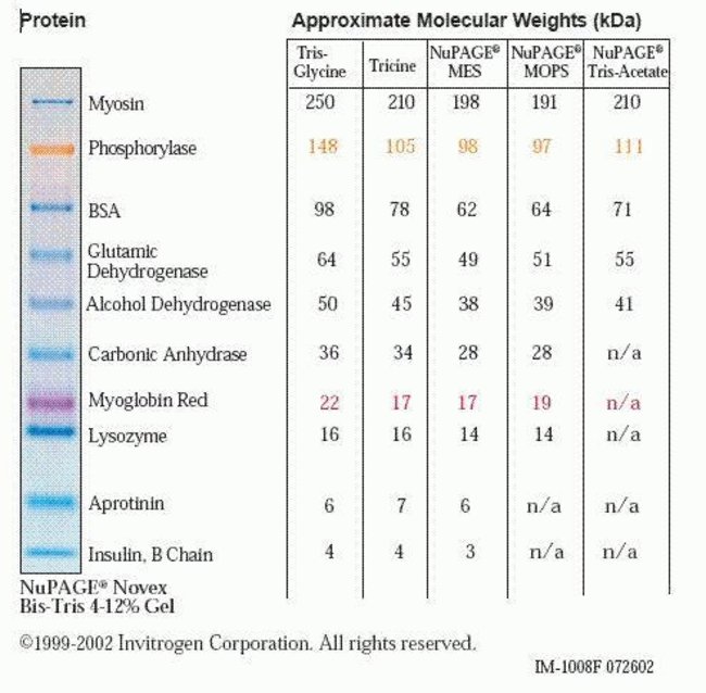
Find additional tips, troubleshooting help, and resources within our Protein Standards and Ladders Support Center.

Can I get the concentration of each protein in the SeeBlue Plus2 Pre-stained Protein Standard (Cat. No. LC5925)?

SeeBlue Plus2 Pre-Stained Standard includes 10 recombinant proteins, that resolve in the range of 4 to 250 kDa depending upon the buffer system being used. Please note that this pre-stained protein standard is not designed for quantitation and we do not recommend that you use this standard in the determination of protein concentration.
Estimations of the protein concentration for SeeBlue Plus2 is given below. However, these values are an approximation and should not be used to quantify samples.
Band per 10 µL: Myosin - 2.20 µg; Phosphorylase-b no estimation is available; BSA - 0.75 µg; GDH - 1.25 µg; Alcohol Dehydrogenase - 0.80 µg; Carbonic Anhydrase- 0.90 µg; Myoglobin (blue)* no estimation available; Lysozyme - 2.50 µg; Aprotinin - 1.80 µg and Insulin - 2.50 µg.

Find additional tips, troubleshooting help, and resources within our Protein Electrophoresis and Western Blotting Support Center.

I used one of your pre-stained protein standards for a western transfer and I noticed that the intensity of the band faded from the membrane during the transfer process. Why is this?

The fading is most likely due to detergent in the western blocking/washing solutions that can remove some of the proteins from the membrane. The dye itself will not wash off of the proteins because it is covalently bound. We have found that smaller pore size membranes retain the proteins better during blocking and wash procedures, and hence recommend use of 0.2 µm instead of 0.45 µm membranes for best resolution and protein retention. After transfer, it is a good idea to circle the pre-stained bands with a pencil on the membrane, so band positions can be identified after blocking and processing.

Find additional tips, troubleshooting help, and resources within our Protein Assays and Analysis Support Center.