Search


Citas y referencias (8)
Invitrogen™
HCS CellMask™ Stains
Easily visualize entire cells or individual cell structures during high-content screening (HCS) assays with HCS CellMask stains, which are available in a variety of colors for multiplexing flexibility and can be used immediately after fixation and permeabilization, or after antibody labeling.
Have Questions?
Cambiar vista

| Número de catálogo | Descripción |
|---|---|
| H32713 | Tinción HCS CellMask™ Orange |
| H32720 | Tinción HCS CellMask Blue |
| H32714 | Tinción HCS CellMask™ Green |
| H32722 | Tinción cercana a IR HCS CellMask™ |
| H32712 | Tinción HCS CellMask™ Red |
| H32721 | Tinción HCS CellMask™ Deep Red |
Número de catálogo H32713
Precio (MXN)
-
Descripción:
Tinción HCS CellMask™ Orange
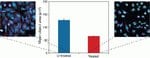
Identify entire cells (cytoplasm and nucleus) during high-content screening (HCS) assays with the versatile HCS CellMask Red, CellMask Orange, CellMask Green, CellMask Blue, CellMask Deep Red, and CellMask Near-IR stains, which are fluorophores that can be used immediately after fixation and permeabilization, or after antibody labeling. They are available in a variety of colors for multiplexing protocols.
The versatile HCS CellMask Red, CellMask Orange, CellMask Green, CellMask Blue, CellMask Deep Red, and CellMask Near-IR stains are cell delineation tools for high content screening (HCS) platforms, label the entire cell (cytoplasm and nucleus) and provide an accurate backdrop against which the features of interest can be assessed. The HCS CellMask Red and Blue stains replace the HCS CellMask Red cytoplasmic/nuclear (Cat. No. H32711) and HCS CellMask Blue cytoplasmic/nuclear stains (Cat. No. H34558), respectively.
HCS CellMask stains can be applied to cells immediately after fixation or in the last step of multiplexing protocols, and they are compatible with detergent-based cell permeabilization protocols. If only nuclear staining is desired, HCS NuclearMask stains can be used for measuring DNA content in addition to enabling robust cell demarcation. HCS NuclearMask stains are also available in a choice of colors for multiplexing flexibility.
HCS CellMask stains can be applied to cells immediately after fixation or in the last step of multiplexing protocols, and they are compatible with detergent-based cell permeabilization protocols. If only nuclear staining is desired, HCS NuclearMask stains can be used for measuring DNA content in addition to enabling robust cell demarcation. HCS NuclearMask stains are also available in a choice of colors for multiplexing flexibility.
Para uso exclusivo en investigación. No apto para uso diagnóstico o terapéutico en humanos ni en animales.
Especificaciones
ColorNaranja
DescripciónTinción HCS CellMask™ Orange
Método de detecciónFluorescente
EmisiónVisible
Intervalo de longitud de onda de excitación556/572
Para utilizar con (equipo)Instrumentos de alto contenido
Línea de productosCellMask
Cantidad1 juego
Condiciones de envíoTemperatura ambiente
Tipo de etiquetaColorante fluorescente
Tipo de productoTinción
Sub Cellular LocalizationNúcleo, citoplasma y citosol, Cytosol, Nucleus
Unit SizeEach
Contenido y almacenamiento
Incluye 1 juego de viales, que proporciona suficiente material para teñir un total de diez microplacas de 96 pocillos
Almacenar a ≤ -20 °C, disecar y proteger de la luz.
Almacenar a ≤ -20 °C, disecar y proteger de la luz.
Preguntas frecuentes
What dyes are used to make the CellMask stains?
Citations & References (8)
Citations & References
Abstract
The Amino Acid Transporter Mct10/Tat1 Is Important to Maintain the TSH Receptor at Its Canonical Basolateral Localization and Assures Regular Turnover of Thyroid Follicle Cells in Male Mice.
Journal:Int J Mol Sci
PubMed ID:34071318
The Thyroid Hormone Transporter Mct8 Restricts Cathepsin-Mediated Thyroglobulin Processing in Male Mice through Thyroid Auto-Regulatory Mechanisms That Encompass Autophagy.
Journal:Int J Mol Sci
PubMed ID:33466458
SLIT2/ROBO2 signaling pathway inhibits nonmuscle myosin IIA activity and destabilizes kidney podocyte adhesion.
Journal:JCI Insight
PubMed ID:27882344
Defining the Cardiac Fibroblast Secretome in a Fibrotic Microenvironment.
Journal:J Am Heart Assoc
PubMed ID:32924724
Canonical TSH Regulation of Cathepsin-Mediated Thyroglobulin Processing in the Thyroid Gland of Male Mice Requires Taar1 Expression.
Journal:Front Pharmacol
PubMed ID:29615904