Search


Citas y referencias (35)
Invitrogen™
pHrodo™ BioParticles™ Conjugates for Phagocytosis and Phagocytosis Kit, for Flow Cytometry
Obtenga resultados más rápidos y exactos en ensayos de fagocitosis con los conjugados y el kit pHrodo BioParticles. Los conjugados pHrodo BioParticles muestran una fluorescencia brillante en los endosomas y lisosomas, no requieren pasos de lavado ni colorantes supresores, se pueden multiplexar y se utilizan en citometría de flujo, HCA y HTS.
Have Questions?
Cambiar vista

| Número de catálogo | Tipo de producto | Especie | Color |
|---|---|---|---|
| P35361 | Conjugado | E. coli | |
| P35360 | Conjugate | E. coli | |
| A10025 | Kit de fagocitosis | E. coli | |
| A10010 | Conjugado | S. aureus | |
| P35367 | Conjugado | S. aureus | |
| P35365 | Conjugado | S. cerevisiae |
Número de catálogo P35361
Precio (MXN)
-
Tipo de producto:
Conjugado
Especie:
E. coli
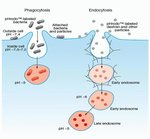
Consiga una tinción más rápida y resultados más exactos sin necesidad de pasos de lavado o colorantes supresores con conjugados pHrodo BioParticles sensibles al pH y kit de fagocitosis para citometría de flujo. Los conjugados pHrodo BioParticles para fagocitosis no son fluorescentes fuera de la celda con pH neutro, pero muestran fluorescencia brillante en ambientes con pH ácido, como los de los endosomas y lisosomas. Se pueden utilizar en aplicaciones de adquisición de imágenes, HCA y HTA. El kit de fagocitosis pHrodo BioParticles ofrece una evaluación rápida de la actividad fagocítica en sangre completa mediante citometría de flujo.
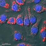
The fluorescence of pHrodo dye dramatically increases as pH decreases from neutral to acidic, making it an ideal tool to study phagocytosis and its regulation by drugs and/or environmental factors. Outside the cell, the dye is non-fluorescent, which eliminates the need for wash steps and quencher dyes. Once internalized, the dye fluoresces brightly within the acidic environments of endosomes and lysosomes, enabling rapid assay development and certainty of results when investigating phagocytic pathways and their regulation by drugs and/or environmental factors. As such, pHrodo dye conjugates can be used in plate readers, fluorescence microscopy imaging, and flow cytometry applications.
The pHrodo Red S. aureus BioParticles Conjugate for Phagocytosis (Cat. No. A10010) offers fast and accurate assay results. pHrodo Red dye conjugates are non-fluorescent outside the cell but fluoresce bright red in phagosomes. The pHrodo Red E. coli BioParticles Conjugate for Phagocytosis (Cat. No. P35361) detects phagocytosis and endocytosis, offers reduced signal variability and improved timing in sensitive experiments, and can be used in multiplex applications with a wide variety of blue, green, and far-red dyes and reporters such as GFP, Fuo-4, calcein, NucBlue, CellEvent Caspase 3/7 green, Mitosox Green, and Mitotracker Deep Red, among many others. The optimal absorption and fluorescence emission maxima of the pHrodo Red BioParticles Conjugate is approximately 560/585 nm, respectively. However, the fluorophore is readily excited with the 488-nm argon-ion laser installed on most flow cytometers.
The pHrodo Red E. coli BioParticles Phagocytosis Kit (Cat. No. A10025) for flow cytometry is designed for rapid and convenient measurement of phagocytic activity in whole blood samples by flow cytometry. The kit includes all the reagents required for assessing particle ingestion and red blood cell lysis.
pHrodo Deep Red dye is a low-background pH sensor dye that shows no signal in neutral conditions and only fluoresces in acidic environments. pHrodo Deep Red dye enables better discrimination of internalized cargo from outside the cell because it has an approximate pKa of 5 and does not fluoresce until it enters the late endosome and lysosome. pHrodo Deep Red dye can be detected using a Cy5 fluorescent filter set and has been validated for use in a variety of applications, including flow cytometry, fluorescent microscopy, high content analysis (HCA), and high throughput screening (HTS). It can also be multiplexed with a wide variety of blue, green, and red dyes and reporters such as GFP and RFPs, Mitosox Green or Red, CellEvent Caspase 3/7 Green or Red, NucBlue, or TMR, among many others.
pHrodo Deep Red E. coli BioParticles Conjugate for Phagocytosis (Cat. No. P35360) is a no-wash, low background, fluorogenic reagent developed for the study of phagocytosis in a live cell system. This conjugate is non-fluorescent outside the cell but fluoresces dark red in phagosomes. The low pKa of the pHrodo Deep Red E. coli BioParticles Conjugate eliminates non-specific signal from non-internalized cargo and only turns on in late endosomes and lysosomes.
The pHrodo Green dye conjugates, like pHrodo Red and pHrodo Deep Red dye conjugates, offer faster and more accurate results than any other phagocytosis assay. pHrodo Green conjugates are non-fluorescent outside the cell at neutral pH but fluoresce bright green at acidic pH, such as that of phagosomes. Use the ready-made pHrodo Green S. aureus BioParticles Conjugate (Cat. No. P35367) and pHrodo Green Zymosan BioParticles Conjugate (Cat. No. P35365) for Phagocytosis in imaging, HCA, HTS, and flow applications.
These pHrodo conjugate products include enough reagent to perform 100 tests when using 100 μL in each well of a 96-well plate, and they can be multiplexed with a wide variety of blue, red, and far-red dye reporters such as Mitosox Red, CellEvent Caspase 3/7 Red, NucBlue, RFPs, and Mitotracker Deep Red, among many others.
pHrodo Red Zymosan BioParticles Conjugate (Cat. No. P35364) is also available, and pHrodo Green dye is available as a conjugate of E. coli BioParticles (Cat. No. P35366).
For Research Use Only. Not for human or animal therapeutic or diagnostic use.
Especificaciones
DescripciónConjugado para fagocitosis pHrodo™ Red-E. coli BioParticles™
Tipo de colorantepHrodo™ Red
FormularioSólido
Cantidad5 x 2 mg
Condiciones de envíoTemperatura ambiente
EspecieE. coli
ColorRojo
Para utilizar con (aplicación)Análisis celular
Para utilizar con (equipo)Microscopio de fluorescencia
Línea de productosBioParticles, pHrodo
Tipo de productoConjugado
Unit SizeEach
Contenido y almacenamiento
Almacenar en el congelador (de –5 °C a –30 °C) y proteger de la luz.
Preguntas frecuentes
I am performing a phagocytosis assay of macrophages engulfing pHrodo-labeled bacteria. What do you recommend for fixation after the phagocytosis?
Can I store reconstituted pHrodo BioParticles Conjugates for Phagocytosis and Phagocytosis Kit, for Flow Cytometry?
Are the Invitrogen BioParticles products sterile?
What is the concentration of bacterial particles for pHrodo BioParticles Conjugates for Phagocytosis?
What is the type of bond that attaches the dyes to the BioParticles probes?
Citations & References (35)
Citations & References
Abstract
Extracellular Matrix Lumican Promotes Bacterial Phagocytosis, and Lum-/- Mice Show Increased Pseudomonas aeruginosa Lung Infection Severity.
Journal:J Biol Chem
PubMed ID:22865855
'Phagocytosis is central to bacterial clearance, but the exact mechanism is incompletely understood. Here, we show a novel and critical role for lumican, the connective tissue extracellular matrix small leucine-rich repeat proteoglycan, in CD14-mediated bacterial phagocytosis. In Psuedomonas aeruginosa lung infections, lumican-deficient (Lum(-/-)) mice failed to clear the bacterium from
SLAM is a microbial sensor that regulates bacterial phagosome functions in macrophages.
Journal:Nat Immunol
PubMed ID:20818396
'Phagocytosis is a pivotal process by which macrophages eliminate microorganisms after recognition by pathogen sensors. Here we unexpectedly found that the self ligand and cell surface receptor SLAM functioned not only as a costimulatory molecule but also as a microbial sensor that controlled the killing of gram-negative bacteria by macrophages.
Innate immunity and transcription of MGAT-III and Toll-like receptors in Alzheimer's disease patients are improved by bisdemethoxycurcumin.
Journal:Proc Natl Acad Sci U S A
PubMed ID:17652175
'We have tested a hypothesis that the natural product curcuminoids, which has epidemiologic and experimental rationale for use in AD, may improve the innate immune system and increase amyloid-beta (Abeta) clearance from the brain of patients with sporadic Alzheimer''s disease (AD). Macrophages of a majority of AD patients do not
Establishment of single-cell screening system for the rapid identification of transcriptional modulators involved in direct cell reprogramming.
Journal:Nucleic Acids Res
PubMed ID:22879381
'Combinatorial interactions of transcription modulators are critical to regulate cell-specific expression and to drive direct cell reprogramming (e.g. trans-differentiation). However, the identification of key transcription modulators from myriad of candidate genes is laborious and time consuming. To rapidly identify key regulatory factors involved in direct cell reprogramming, we established a
Rooperol as an antioxidant and its role in the innate immune system: An in vitro study.
Journal:J Ethnopharmacol
PubMed ID:23085395
'Biologically active rooperol is formed when the glucose subunits of the nontoxic glycoside, hypoxoside, are cleaved by ß-glucosidase. Hypoxoside is isolated from Hypoxis, a medicinal plant genus frequently used by the indigenous people of South Africa as an immune system booster. The aim of this study was to investigate rooperol''s