Search


引用資料與參考文獻 (30)
Invitrogen™
ProLong™ Gold Antifade Mountant with DNA Stain DAPI
ProLong Gold Antifade Mountant with DNA Stain DAPI come mixed and ready-to-use to make counterstaining more convenient and consistent while protecting fluorescently stained samples.
Have Questions?
變更視圖

| 產品號碼 | Quantity |
|---|---|
| P36935 | 5 x 2 mL |
| P36941 | 1 x 2 mL |
| P36931 | 1 x 10 mL |
產品號碼 P36935
價格 (TWD)
10,560.00
線上優惠
Ends: 30-Jun-2026
13,200.00您節省 2,640.00 (20%)
Each
Quantity:
5 x 2 mL
價格 (TWD)
10,560.00
線上優惠
Ends: 30-Jun-2026
13,200.00您節省 2,640.00 (20%)
Each
ProLong Gold Antifade Mountant with DNA Stain DAPI is a mounting media that can be directly applied to fluorescently labeled cell or tissue samples on microscope slides. It comes ready-to-use and stored at room temperature—just apply a drop, add a coverslip, cure, and image. ProLong Gold Antifade Mountants come in a 2-mL dropper bottle or 10-mL bottle. It is formulated with the blue DNA stain DAPI to make your nuclear counterstaining more convenient and consistent when preparing your samples for imaging.
ProLong Gold Antifade Mountant with DNA Stain DAPI contains chemical components designed to protect fluorescent dyes from fading (photobleaching) during fluorescence microscopy experiments. It is a curing mounting media that forms an optical path of 1.47 RI and allows longer-term storage of the sample.
Many mountants can quench initial fluorescence signal strength by up to 80%. ProLong Gold Antifade Mountant was developed to minimize this initial signal quenching for the broadest range of colors and dyes and is formulated with the DAPI DNA nuclear stain to simplify your experiments. DAPI stain is excited by UV light at 360 nm when bound to DNA, with an emission maximum at 460 nm, and is detected using a DAPI traditional filter.
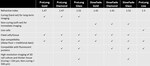
ProLong Gold Antifade Mountant is not recommended for mounting samples containing fluorescent proteins such as GFP or TagRFP but can be used with Alexa Fluor dyes of your choice. For superior antifade protection of fluorescent proteins and fluorescent dyes, ProLong Diamond or ProLong Glass Antifade Mountants are recommended. ProLong Glass mountants are recommended for all immersion oil imaging applications. For immediate viewing of a sample, choose our non-curing mountant, SlowFade Gold Antifade Mountant with DAPI.
For Research Use Only. Not for use in diagnostic procedures.
規格
Quantity5 x 2 mL
Shipping ConditionRoom Temperature
Product LineProLong
Product TypeAntifade Mountant
Reagent TypeMounting Solution, Antifade Solution
Volume (Metric)2 mL
Unit SizeEach
內容物與存放
Storage at room temperature is recommended but can also be stored frozen (-5 to -30°C). Protect from light.
常見問答集 (常見問題)
What is the difference between ProLong and SlowFade antifade reagents?
If I use ProLong Gold Antifade Mountant to mount my slides, should I seal the edges?
Some antifade mounting media stay as liquid whereas others harden. What is the benefit of having one that hardens?
I mounted my cells in ProLong antifade mounting medium, but now I want to go back and re-label them. Is there a way I can unmount the coverslip after it has cured (hardened)?
I am using ProLong antifade mounting medium. Do I need to let it cure before imaging? Do I need to seal the edges of the coverslip?
引用資料與參考文獻 (30)
引用資料與參考文獻
Abstract
Super-resolution imaging reveals three-dimensional folding dynamics of the ß-globin locus upon gene activation.
Journal:J Cell Sci
PubMed ID:22767512
The chromatin architecture is constantly changing because of cellular processes such as proliferation, differentiation and changes in the expression profile during gene activation or silencing. Unravelling the changes that occur in the chromatin structure during these processes has been a topic of interest for many years. It is known that
Resolution of de novo HIV production and trafficking in immature dendritic cells.
Journal:Nat Methods
PubMed ID:18059278
'The challenge in observing de novo virus production in human immunodeficiency virus (HIV)-infected dendritic cells (DCs) is the lack of resolution between cytosolic immature and endocytic mature HIV gag protein. To track HIV production, we developed an infectious HIV construct bearing a diothiol-resistant tetracysteine motif (dTCM) at the C terminus
Androgen induces expression of the multidrug resistance protein gene MRP4 in prostate cancer cells.
Journal:Prostate Cancer Prostatic Dis
PubMed ID:17003774
'Multidrug resistance-associated proteins (MRPs) may mediate multidrug resistance in tumor cells. Using a gene array analysis, we have identified MRP4 as an androgen receptor (AR)-regulated gene. Dihydrotestosterone induced MRP4 expression in both androgen-dependent and -independent LNCaP cells, whereas there was little detectable expression in PC-3 or normal prostate epithelial cells.
H2AX phosphorylation within the G1 phase after UV irradiation depends on nucleotide excision repair and not DNA double-strand breaks.
Journal:Proc Natl Acad Sci U S A
PubMed ID:16788066
'The variant histone H2AX is phosphorylated in response to UV irradiation of primary human fibroblasts in a complex fashion that is radically different from that commonly reported after DNA double-strand breaks. H2AX phosphorylation after exposure to ionizing radiation produces foci, which are detectable by immunofluorescence microscopy and have been adopted
AZD2171: a highly potent, orally bioavailable, vascular endothelial growth factor receptor-2 tyrosine kinase inhibitor for the treatment of cancer.
Journal:Cancer Res
PubMed ID:15899831
'Inhibition of vascular endothelial growth factor-A (VEGF) signaling is a promising therapeutic approach that aims to stabilize the progression of solid malignancies by abrogating tumor-induced angiogenesis. This may be accomplished by inhibiting the kinase activity of VEGF receptor-2 (KDR), which has a key role in mediating VEGF-induced responses. The novel