
Webinar
Webinar | Quantifying metal ageing to build more durable components
In this webinar, we will explore how to validate the mechanical performance of metals and alloys and how to utilize them to produce reliable, long-lasting
In this webinar, we will explore how to validate the mechanical performance of metals and alloys and how to utilize them to produce reliable, long-lasting

While FIB-SEM generates incredibly detailed 3D datasets, extracting meaningful insights from complex ultrastructure can require extensive manual segmentation and quantification. Amira Software streamlines this process

InterPore 2026 is the 18th Annual Meeting of the International Society for Porous Media (InterPore), a major scientific conference focused on porous media research. Things

Join our live webinar to see how AI-enabled workflows in Amira Software can streamline your analysis from image enhacement, segmentation to data interpretation, and help

VIZBI is an international conference on visualizing complex biological data, uniting scientists, designers, and artists.It fosters collaboration and shares tools, methods, and best practices for

The Youth Geoscience Forum was initiated by Chinese young geoscientists. It is led by renowned scientists as advisors and consists of young scholars and graduate

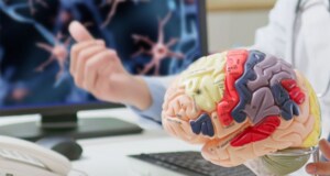
Our new eBook showcases how Thermo Scientific™ Amira™ Software supports advanced neuroscience research. While modern neuroscience imaging delivers extraordinary detail across scales, turning complex, high-resolution

Xtras are add-ons and how-to videos that will help you improve your daily use of Amira and Avizo Software. Add on: ML/DL Object Classification and

Join our upcoming webinar to learn how advanced electron microscopy and image analysis can help you quantify the nanoscale properties that drive catalytic performance. Discover
Light-sheet microscopy combined with tissue clearing and expansion offers a nondestructive way to image neural tissue in true 3D without mechanical sectioning, preserving structural integrity