Search
Zeta
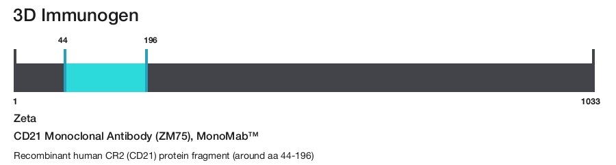
CD21 Monoclonal Antibody (ZM75), MonoMab™
{{$productOrderCtrl.translations['antibody.pdp.commerceCard.promotion.promotions']}}
{{$productOrderCtrl.translations['antibody.pdp.commerceCard.promotion.viewpromo']}}
{{$productOrderCtrl.translations['antibody.pdp.commerceCard.promotion.promocode']}}: {{promo.promoCode}} {{promo.promoTitle}} {{promo.promoDescription}}. {{$productOrderCtrl.translations['antibody.pdp.commerceCard.promotion.learnmore']}}
Additional Information:
{{banner.description}}
 (IHC (P))")

 in IHC (P)")
Product Details
Z2385MS
Product Specifications
Species Reactivity
Human
Host/Isotype
Mouse
/ IgG1, kappa
Class
Monoclonal
Type
Antibody
Clone
ZM75
Immunogen
Recombinant human CR2 (CD21) protein fragment (around aa 44-196)
Conjugate
Form
Liquid
Concentration
200 µg/mL
Amount
100 µg
Purification
Protein A
Storage buffer
tris with BSA, NP-40
Contains
<0.1% sodium azide
Storage conditions
4°C
Shipping conditions
Ambient (domestic); Wet ice (international)
Product Specific Information
A recommended positive control tissue for this product is Lymph Node, however positive controls are not limited to this tissue type.
The primary antibody is intended for laboratory professional use in the detection of the corresponding protein in formalin-fixed, paraffin-embedded tissue stained in manual qualitative immunohistochemistry (IHC) testing. This antibody is intended to be used after the primary diagnosis of tumor has been made by conventional histopathology using non-immunological histochemical stains.
Recognizes a protein of 140 kDa, which is identified as the complement receptor 2 (CR2)/CD21. This protein is expressed strongly on mature B cells, follicular dendritic cells and weakly on immature thymocytes and T lymphocytes. In B-cell ontogeny, CD21 appears after the pre-B-stage, is maintained during peripheral B-cell development and is lost upon terminal differentiation into plasma cells. CD21 expression is also gradually lost after stimulation of B cells in vitro. CD21 functions as receptor for C3d, C3dg and iC3b Complement components, for EBV and for IFN alpha. CD21 binds to CD23 and associates with CD19, CD81 and Leu13 to form a large signal-transduction complex involved in B cell activation.
Antibody is used with formalin-fixed and paraffin-embedded sections. Pretreatment of deparaffinized tissue with heat-induced epitope retrieval or enzymatic retrieval is recommended. In general, immunohistochemical (IHC) staining techniques allow for the visualization of antigens via the sequential application of a specific antibody to the antigen (primary antibody), a secondary antibody to the primary antibody (link antibody), an enzyme complex and a chromogenic substrate with interposed washing steps. The enzymatic activation of the chromogen results in a visible reaction product at the antigen site. Results are interpreted using a light microscope and aid in the differential diagnosis of pathophysiological processes, which may or may not be associated with a particular antigen.
A positive tissue control must be run with every staining procedure performed. This tissue may contain both positive and negative staining cells or tissue components and serve as both the positive and negative control tissue. External Positive control materials should be fresh autopsy/biopsy/surgical specimens fixed, processed and embedded as soon as possible in the same manner as the patient sample (s). Positive tissue controls are indicative of correctly prepared tissues and proper staining methods. The tissues used for the external positive control materials should be selected from the patient specimens with well-characterized low levels of the positive target activity that gives weak positive staining. The low level of positivity for external positive controls is designed to ensure detection of subtle changes in the primary antibody sensitivity from instability or problems with the staining methodology. A tissue with weak positive staining is more suitable for optimal quality control and for detecting minor levels of reagent degradation.
Internal or external negative control tissue may be used depending on the guidelines and policies that govern the organization to which the end user belongs to. The variety of cell types present in many tissue sections offers internal negative control sites, but this should be verified by the user. The components that do not stain should demonstrate the absence of specific staining, and provide an indication of non-specific background staining. If specific staining occurs in the negative tissue control sites, results with the patient specimens must be considered invalid.
Target Information
CD21 (complement receptor 2, CR2, C3D receptor, EBV receptor) binds C3 complement fragments, especially its breakdown fragments, which remain covalently attached to complement activating surfaces or antigen. CD21 has important roles in uptake and retention of immunocomplexes, survival of memory B cells and in development and maintenance of the humoral response to T-dependent antigens. CD21 also serves as a key receptor for Epstein-Barr virus binding and is involved in targeting prions to follicular dendritic cells and expediting neuroinvasion following peripheral exposure to prions. A soluble form of the CD21 (sCD21) is shed from the lymphocyte surface and retains its ability to bind respective ligands. CD21 functions as receptor for C3d, C3dg and iC3b complement components, for EBV and for IFNalpha. CD21 binds to CD23 and associates with CD19, CD81 and Leu13 to form a large signal-transduction complex involved in B cell activation. Genetic variations in the CD21 gene are associated with susceptibility to systemic lupus erythematosus type 9 (SLEB9). Alternatively, spliced transcript variants encoding different isoforms of CD21 have been found.
For Research Use Only. Not for use in diagnostic procedures. Not for resale without express authorization.
References (0)
Have you cited this product in a publication?
Let us know so we can reference it here.
Bioinformatics
Protein Aliases: CD21; Complement C3d receptor; Complement C3d receptor (C3DR); complement component (3d/Epstein Barr virus) receptor 2; complement component 3d receptor 2; Complement receptor type 2; Complement Receptor type 2 (CR2); Cr2; CR2 precursor; CR2/CD21/C3d/Epstein-Barr virus receptor precursor; EBV receptor; EBV-R; Epstein-Barr virus receptor; EVBR; unnamed protein product
Gene Aliases: C3DR; CD21; CR; CR2; CVID7; SLEB9
UniProt ID: (Human) P20023
Entrez Gene ID: (Human) 1380
immune system process
complement receptor mediated signaling pathway
T cell mediated immunity
immune response
complement activation, alternative pathway
complement activation, classical pathway
B cell differentiation
B cell proliferation
B cell activation
innate immune response
negative regulation of complement activation, classical pathway
viral entry into host cell
type I interferon signaling pathway
antiviral innate immune response
Disclaimer

Clicking the images or links will redirect you to a website hosted by BenchSci that provides third-party scientific content. Neither the content nor the BenchSci technology and processes for selection have been evaluated by us; we are providing them as-is and without warranty of any kind, including for use or application of the Thermo Fisher Scientific products presented.

Performance Guarantee
If an Invitrogen™ antibody doesn't perform as described on our website or datasheet,we'll replace the product at no cost to you, or provide you with a credit for a future purchase.*
Learn more
We're here to help
Get expert recommendations for common problems or connect directly with an on staff expert for technical assistance related to applications, equipment and general product use.
Contact tech support