Search Thermo Fisher Scientific
Disclaimer

Clicking the images or links will redirect you to a website hosted by BenchSci that provides third-party scientific content. Neither the content nor the BenchSci technology and processes for selection have been evaluated by us; we are providing them as-is and without warranty of any kind, including for use or application of the Thermo Fisher Scientific products presented.
Invitrogen
CD279 (PD-1) Monoclonal Antibody (J116), eBioscience™
 Antibody in Flow Cytometry (Flow)")
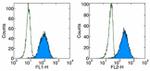


 Antibody (14-9989-82) in Flow")
 Antibody (14-9989-82) in IHC (F)")
 Antibody (14-9989-82) in IHC, IHC (F)")
 Antibody (14-9989-82) in IHC, IHC (F)")
 Antibody (14-9989-82) in IHC")
 Antibody (14-9989-82) in Flow")
 Antibody (14-9989-82) in Flow")
 Antibody (14-9989-82) in Flow")
Product Details
14-9989-82
Applications
Tested Dilution
Publications
Product Specifications
Species Reactivity
Human
Published species
Human
Host/Isotype
Mouse
/ IgG1, kappa
Class
Monoclonal
Type
Antibody
Clone
J116
Conjugate
Unconjugated
Form
Liquid
Concentration
0.5 mg/mL
Purification
Affinity chromatography
Storage buffer
PBS, pH 7.2
Contains
0.09% sodium azide
Storage conditions
4° C
Shipping conditions
Wet ice
RRID
AB_468672
Product Specific Information
Description: The J116 monoclonal antibody reacts with the human PD-1 (programmed death-1), a 55 kDa member of the immunoglobulin superfamily. PD-1 contains the immunoreceptor tyrosine-based inhibitory motif (ITIM) and plays a key role in peripheral tolerance and autoimmune disease. PD-1 is expressed predominantly on activated T and B lymphocytes. Two novel members of the B7 family have been identified as the PD-1 ligands, PD-L1 (B7-H1) and PD-L2 (B7-DC). Evidence reported to date suggests overlapping functions for these two PD-1 ligands and their constitutive expression on some normal tissues and upregulation on activated antigen-presenting cells. Binding of the J116 monoclonal antibody inhibits PD-1 signal transduction, however, it does not block binding of the ligand PD-L1.
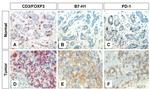
Applications Reported: This J116 antibody has been reported for use in immunoprecipitation and immunohistology staining of frozen tissue sections. It has also been reported in in vitro functional assays. (Please use Functional Grade purified J116, Product # 16-9989 , in functional assays.).
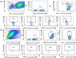
Applications Tested: The J116 antibody has been tested by flow cytometric analysis of human PD-1 transfected cells. This can be used at less than or equal to 1 µg per test. A test is defined as the amount (µg) of antibody that will stain a cell sample in a final volume of 100 µL. Cell number should be determined empirically but can range from 10^5 to 10^8 cells/test. It is recommended that the antibody be carefully titrated for optimal performance in the assay of interest.
Purity: Greater than 90%, as determined by SDS-PAGE.
Aggregation: Less than 10%, as determined by HPLC.
Filtration: 0.2 µm post-manufacturing filtered.
Target Information
Cell-mediated immune responses are initiated by T lymphocytes that are themselves stimulated by cognate peptides bound to MHC molecules on antig en-presenting cells (APC). T-cell activation is generally self-limited as activated T cells express receptors such as PD-1 (also known as PDCD-1) that mediate inhibitory signals from the APC. PD-1 can bind two different but related ligands, PDL-1 and PDL-2. Upon binding to either of these ligands, signals generated by PD-1 inhibit the activation of the immune response in the absence of "danger signals" such as LPS or other molecules associated with bacteria or other pathogens. Evidence for this is seen in PD1-null mice who exhibit hyperactivated immune systems and autoimmune diseases. Despite its predicted molecular weight, PD-1 often migrates at higher molecular weight in SDS-PAGE.
For Research Use Only. Not for use in diagnostic procedures. Not for resale without express authorization.
Bioinformatics
Protein Aliases: CD279; hPD1; programmed cell death 1 protein; Programmed cell death protein 1; Protein PD-1; Protein PD1; sCD279; soluble CD279; systemic lupus erythematosus susceptibility 2
Gene Aliases: CD279; hPD-1; hPD-l; hSLE1; PD-1; PD1; PDCD1; SLEB2
UniProt ID: (Human) Q15116
Entrez Gene ID: (Human) 5133

Performance Guarantee
If an Invitrogen™ antibody doesn't perform as described on our website or datasheet,we'll replace the product at no cost to you, or provide you with a credit for a future purchase.*
Learn more
We're here to help
Get expert recommendations for common problems or connect directly with an on staff expert for technical assistance related to applications, equipment and general product use.
Contact tech support