Search
Zeta
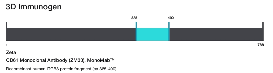
CD61 Monoclonal Antibody (ZM33), MonoMab™
{{$productOrderCtrl.translations['antibody.pdp.commerceCard.promotion.promotions']}}
{{$productOrderCtrl.translations['antibody.pdp.commerceCard.promotion.viewpromo']}}
{{$productOrderCtrl.translations['antibody.pdp.commerceCard.promotion.promocode']}}: {{promo.promoCode}} {{promo.promoTitle}} {{promo.promoDescription}}. {{$productOrderCtrl.translations['antibody.pdp.commerceCard.promotion.learnmore']}}
Additional Information:
{{banner.description}}
Product Details
Z2337MS
Product Specifications
Species Reactivity
Human
Host/Isotype
Mouse
/ IgG2b, kappa
Class
Monoclonal
Type
Antibody
Clone
ZM33
Immunogen
Recombinant human ITGB3 protein fragment (aa 385-490)
Conjugate
Form
Liquid
Concentration
200 µg/mL
Amount
100 µg
Purification
Protein A
Storage buffer
tris with BSA, NP-40
Contains
<0.1% sodium azide
Storage conditions
4°C
Shipping conditions
Ambient (domestic); Wet ice (international)
Product Specific Information
A recommended positive control tissue for this product is Bone marrow, however positive controls are not limited to this tissue type.
The primary antibody is intended for laboratory professional use in the detection of the corresponding protein in formalin-fixed, paraffin-embedded tissue stained in manual qualitative immunohistochemistry (IHC) testing. This antibody is intended to be used after the primary diagnosis of tumor has been made by conventional histopathology using non-immunological histochemical stains.
CD61 (GPIIIa) is a glycoprotein found on megakaryocytes, platelets and their precursors. CD61 antigen plays a role in platelet aggregation and also as a receptor for fibrinogen, fibronectin, von Willebrand factor and vitronectin. Clone 2f2 will prove useful in detecting neoplastic platelet precursors, normal platelets and most cases of megakaryocytic leukemia.
Antibody is used with formalin-fixed and paraffin-embedded sections. Pretreatment of deparaffinized tissue with heat-induced epitope retrieval or enzymatic retrieval is recommended. In general, immunohistochemical (IHC) staining techniques allow for the visualization of antigens via the sequential application of a specific antibody to the antigen (primary antibody), a secondary antibody to the primary antibody (link antibody), an enzyme complex and a chromogenic substrate with interposed washing steps. The enzymatic activation of the chromogen results in a visible reaction product at the antigen site. Results are interpreted using a light microscope and aid in the differential diagnosis of pathophysiological processes, which may or may not be associated with a particular antigen.
A positive tissue control must be run with every staining procedure performed. This tissue may contain both positive and negative staining cells or tissue components and serve as both the positive and negative control tissue. External Positive control materials should be fresh autopsy/biopsy/surgical specimens fixed, processed and embedded as soon as possible in the same manner as the patient sample (s). Positive tissue controls are indicative of correctly prepared tissues and proper staining methods. The tissues used for the external positive control materials should be selected from the patient specimens with well-characterized low levels of the positive target activity that gives weak positive staining. The low level of positivity for external positive controls is designed to ensure detection of subtle changes in the primary antibody sensitivity from instability or problems with the staining methodology. A tissue with weak positive staining is more suitable for optimal quality control and for detecting minor levels of reagent degradation.
Internal or external negative control tissue may be used depending on the guidelines and policies that govern the organization to which the end user belongs to. The variety of cell types present in many tissue sections offers internal negative control sites, but this should be verified by the user. The components that do not stain should demonstrate the absence of specific staining, and provide an indication of non-specific background staining. If specific staining occurs in the negative tissue control sites, results with the patient specimens must be considered invalid.
Target Information
CD61, also known as GPIIIa or ITGB3, is a 105 kDa glycoprotein expressed on activated T cells, granulocytes, megakaryocytes, platelets, and their precursors. It plays a crucial role in platelet aggregation and functions as a receptor for fibrinogen, fibronectin, von Willebrand factor, vitronectin, and thrombospondin. CD61 forms heterodimeric complexes by associating non-covalently with integrin alpha subunits: alphaV (CD51) to create the Vitronectin Receptor and alphaIIb (CD41) to form gpIIb/IIIa. These complexes are responsible for adhesion to extracellular matrix components, facilitating cell adhesion and cell-surface mediated signaling. CD61 is expressed on platelets and megakaryocytes in association with CD41, and on endothelial cells, monocytes, and osteoclasts in association with CD51. The protein product of CD61 is composed of an alpha chain and a beta chain, which can combine with multiple partners to form different integrins. Its involvement in cell adhesion and signaling underscores its importance in normal physiological processes. Dysfunction of CD61 is associated with diseases such as Glanzmann Thrombasthenia and Platelet type-16 Bleeding Disorder, highlighting its critical role in hemostasis and platelet function.
For Research Use Only. Not for use in diagnostic procedures. Not for resale without express authorization.
References (0)
Have you cited this product in a publication?
Let us know so we can reference it here.
Bioinformatics
Protein Aliases: antigen CD61; CD61; glycoprotein IIIa precursor; GPIIIa; integrin beta 3; integrin beta chain, beta 3; Integrin beta-3; integrin beta-3 subunit; integrin, beta 3 (platelet glycoprotein IIIa, antigen CD61); plate glycoprotein IIIa (GPIIIa); Platelet membrane glycoprotein IIIa
Gene Aliases: BDPLT16; BDPLT2; BDPLT24; CD61; GP3A; GPIIIa; GT; GT2; ITGB3
UniProt ID: (Human) P05106
Entrez Gene ID: (Human) 3690
virus receptor activity
fibronectin binding
protease binding
protein disulfide isomerase activity
protein kinase C binding
platelet-derived growth factor receptor binding
integrin binding
protein binding
coreceptor activity
fibroblast growth factor binding
enzyme binding
C-X3-C chemokine binding
insulin-like growth factor I binding
neuregulin binding
identical protein binding
vascular endothelial growth factor receptor 2 binding
metal ion binding
cell adhesion molecule binding
extracellular matrix binding
fibrinogen binding
angiogenesis
positive regulation of endothelial cell proliferation
positive regulation of cell-matrix adhesion
positive regulation of leukocyte migration
cell-substrate junction assembly
cell adhesion
cell-matrix adhesion
integrin-mediated signaling pathway
embryo implantation
blood coagulation
regulation of G-protein coupled receptor protein signaling pathway
positive regulation of cell proliferation
positive regulation of endothelial cell migration
positive regulation of gene expression
negative regulation of macrophage derived foam cell differentiation
positive regulation of fibroblast migration
negative regulation of lipid storage
negative regulation of low-density lipoprotein particle clearance
response to activity
smooth muscle cell migration
positive regulation of smooth muscle cell migration
cell migration
platelet activation
positive regulation of cell migration
positive regulation of vascular endothelial growth factor receptor signaling pathway
cell-substrate adhesion
negative regulation of lipid transport
regulation of protein localization
regulation of actin cytoskeleton organization
cell adhesion mediated by integrin
positive regulation of cell adhesion mediated by integrin
positive regulation of osteoblast proliferation
heterotypic cell-cell adhesion
substrate adhesion-dependent cell spreading
tube development
wound healing, spreading of epidermal cells
response to platelet-derived growth factor
cellular response to platelet-derived growth factor stimulus
apolipoprotein A-I-mediated signaling pathway
wound healing
apoptotic cell clearance
regulation of bone resorption
positive regulation of angiogenesis
positive regulation of bone resorption
viral entry into host cell
platelet-derived growth factor receptor signaling pathway
positive regulation of fibroblast proliferation
mesodermal cell differentiation
positive regulation of smooth muscle cell proliferation
negative regulation of lipoprotein metabolic process
regulation of developmental process
negative chemotaxis
regulation of transport
regulation of release of sequestered calcium ion into cytosol
regulation of serotonin uptake
angiogenesis involved in wound healing
regulation of biological quality
positive regulation of ERK1 and ERK2 cascade
platelet aggregation
cellular response to mechanical stimulus
cellular response to xenobiotic stimulus
positive regulation of glomerular mesangial cell proliferation
blood coagulation, fibrin clot formation
maintenance of postsynaptic specialization structure
regulation of postsynaptic neurotransmitter receptor internalization
regulation of postsynaptic neurotransmitter receptor diffusion trapping
positive regulation of substrate adhesion-dependent cell spreading
positive regulation of adenylate cyclase-inhibiting opioid receptor signaling pathway
positive regulation of vascular endothelial growth factor signaling pathway
regulation of trophoblast cell migration
regulation of extracellular matrix organization
cellular response to insulin-like growth factor stimulus
negative regulation of endothelial cell apoptotic process
positive regulation of T cell migration
Disclaimer

Clicking the images or links will redirect you to a website hosted by BenchSci that provides third-party scientific content. Neither the content nor the BenchSci technology and processes for selection have been evaluated by us; we are providing them as-is and without warranty of any kind, including for use or application of the Thermo Fisher Scientific products presented.

Performance Guarantee
If an Invitrogen™ antibody doesn't perform as described on our website or datasheet,we'll replace the product at no cost to you, or provide you with a credit for a future purchase.*
Learn more
We're here to help
Get expert recommendations for common problems or connect directly with an on staff expert for technical assistance related to applications, equipment and general product use.
Contact tech support