See key cryo-EM structures submitted to the Protein Data Bank on Twitter.
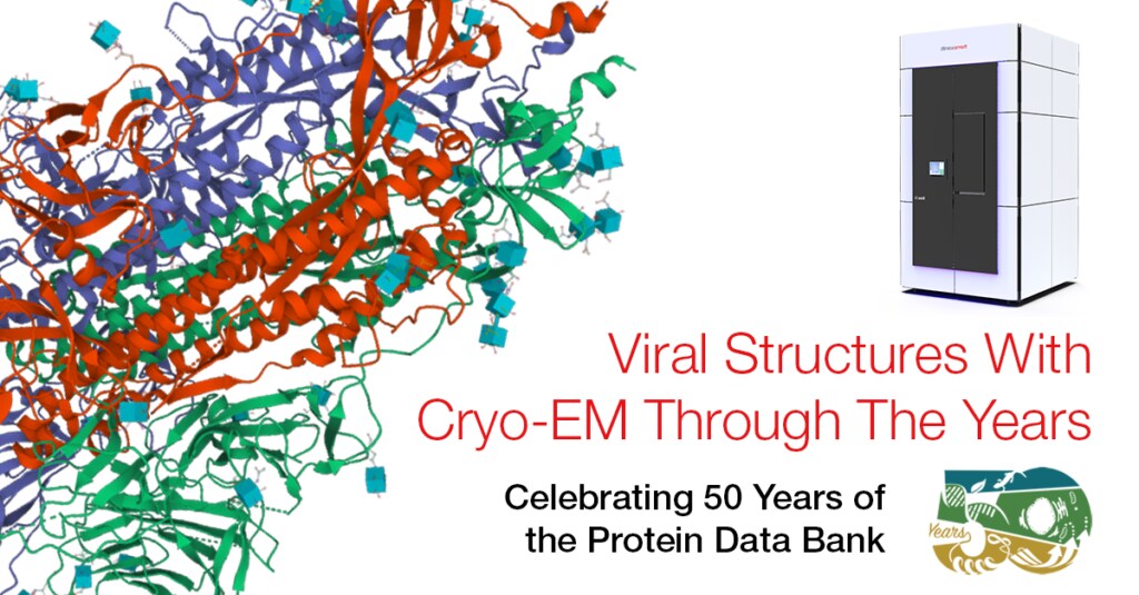
This year, the Protein Data Bank celebrates its 50th anniversary while cryo-electron microscopy (cryo-EM) continues to rise in prominence as the go-to technique for solving biological structures at high-resolution. Thanks in part to increased cryo-EM access, the total number of solved structures added to this single worldwide archive grew fivefold over the past two decades.
In that time, the number of structures added via electron microscopy alone grew by nearly 17,000%. In 2020, 2,390 structures solved using cryo-EM were added to the archive—a 65% jump over the previous year and 15% of all structures submitted that year.
Cryo-EM ushered in a new era of structural biology with its ability to view molecules in their near-native state at atomic-level resolution. The method became the second most widely used technique for solving structures added to the Protein Data Bank, surpassing nuclear magnetic resonance (NMR) in 2017.
Cryo-EM also gained ground on the No. 1 method to-date—X-ray crystallography—which faces limits in its ability to crystallize macromolecules of large size and poor stability.
Cryo-EM access through innovative instrumentation
As cryo-EM continues its ascent, it is important to callout the cutting-edge equipment that made it possible.
Thanks to their speed, accuracy, and high-resolution results, our Thermo Scientific Glacios Cryo-TEM, Krios Cryo-TEM, and Talos Arctica Cryo-TEM contribute to the structures solved using cryo-EM and lead to important discoveries that increase our understanding of a wide range of diseases from HIV to cancer to neurodegenerative diseases like Alzheimer’s or Parkinson’s.
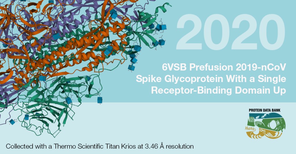
This is the first cryo-EM structure of the SARS-CoV-2 spike protein. The rapid mapping of this structure led to subsequent drug development.
Moreover, related techniques such as cryo-electron tomography (cryo-ET) revolutionized biomedical research by making it possible to visualize 3D protein structures and disease states within the context of the larger cell.
Adopting cryo-EM
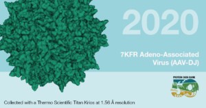
One of the highest resolution structures deposited: adeno-associated virus, the leading viral vector for gene therapy.
While cryo-EM has come a long way, realizing the full potential of this technique will require a far greater level of adoption. Whether it be new diseases and viruses or a changing environment that puts new strains on agricultural crops, far more scientists in many research areas need access to cryo-EM technologies to tackle the evolving set of challenges in front of them.
//
Jeffrey Lengyel, Ph.D., is Principal Scientist of Life Sciences at Thermo Fisher Scientific. To learn more about the breakthrough impacts of cryo-EM, visit our Life Sciences Electron Microscopy Learning Center.

Advances in high-resolution cryo-EM at 100 kV
100 kV cryo-TEM enables high-resolution single particle anal... Alex Ilitchev, PhD
Read More
3D Tissue Histology with Light-Sheet Microscopy Enables Nondestructive Analysis of Microglia
3D tissue analysis offers critical benefits for neuroscience...
Read More
Fragment based drug discovery meets challenging drug targets with high-throughput cryo-EM
Benefits of FBDD in the search for novel therapeutics Frag...
Read More
Targeted protein degradation as a novel therapeutic approach for undruggable diseases
Induced proximity for targeted protein degradation In 1993, ...
Read More
Leave a Reply