Using mouse models of Alzheimer’s disease to discover the distribution and behavior of amyloid plaques
Neurological disorders are an ongoing pressure on the continuously growing elderly population worldwide, with few treatments or even management options available to many. This causes significant economic and social hardship, not just to those afflicted, but to their families and society as a whole. Of these illnesses, Alzheimer’s disease is perhaps one of the most intensely researched, partly because of its distinctive post-mortem pathology, characterized by pompom-like plaques in the brain.
These plaques are composed primarily of amyloid-beta, a functional protein involved in neurological maintenance. In patients with Alzheimer’s disease, this protein misfolds into a non-functional conformation that is highly prone to aggregation, creating strands that eventually bundle together into globular amyloid plaques. The process of amyloid-beta aggregation is closely tied to neurological degeneration, although the exact mechanism is still unknown. Mouse models of Alzheimer’s disease can provide a look at early phases of the disease, before symptoms may even manifest in human patients. This makes them an essential tool in Alzheimer’s research.

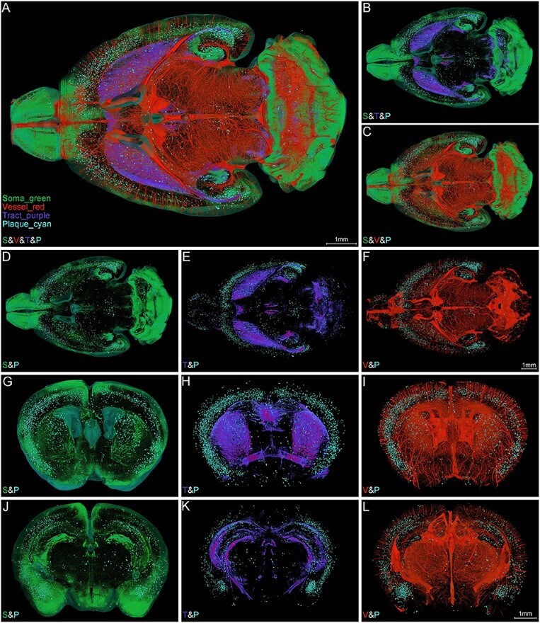
Whole brain mouse model of Alzheimer’s disease produced with micro-optical sectioning tomography and reconstruction in Amira Software. Image reproduced under CC BY 4.0 from X. Yin, et al.
Micro-optical sectioning tomography and an image processing workflow provides unprecedented detail in an Alzheimer’s mouse model
The ultimate goal of these mouse models is a clear understanding of disease progression, leading to treatments that target amyloid aggregation at its root. Whole-brain imaging would inherently provide the most complete picture possible, but is no trivial task, even for the relatively small mouse brain. Between complex sample preparation, high-resolution imaging, and data size/processing, high-quality whole-brain imaging has remained an elusive target.
Researchers at the Huazhong University of Science and Technology recently employed micro-optical sectioning tomography to produce a fluorescence-free whole-brain mouse model of Alzheimer’s disease. Nissl staining provided the contrast needed for structural differentiation. This technology combined with a customized image processing workflow allowed the researchers to visualize the distribution of the Amyloid beta plaques and their spatial correlation with different brain components in the whole brain of Alzheimer disease mice. The image processing workflow -includes- channel splitting, feature enhancement, iso-surface rendering as well volume rendering. This workflow can be also applied in other high-throughput bright field imaging to obtain a comprehensive understanding of the complex brain.

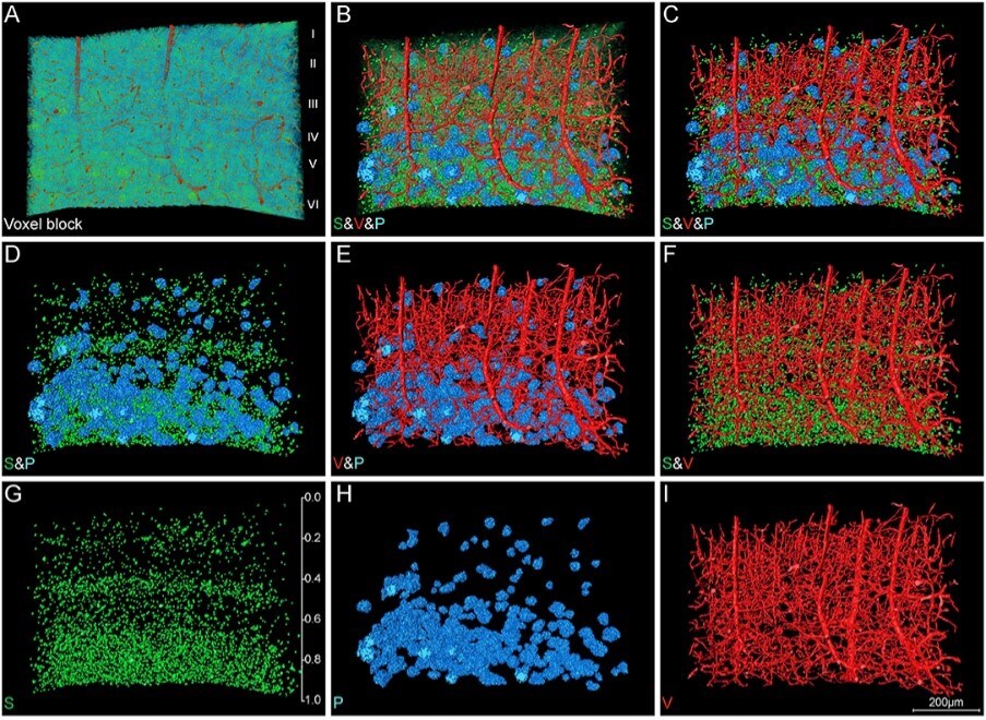
Close up of a mouse brain region containing amyloid plaques. Their distribution and location relative to the blood vessels and nerve cells can be clearly differentiated. (Plaques = blue, soma = green, vessels = red.) Image reproduced under CC BY 4.0 from X. Yin, et al.
Fluorescence-free tomography reconstruction and visualization with Amira Software
Achieving accurate data segmentation and reconstruction can be challenging, especially with large datasets, which is why the researchers turned to Thermo Scientific Amira Software for their data processing. It allowed them to automate some of the repetitive work and even develop custom scripts to replace routine manual tasks, greatly streamlining the overall process. Using specialized tools to generate volume renderings and 3D surfaces in Amira Software, they were able to clearly differentiate blood vessels, amyloid plaques, as well as neuronal soma and tracts. This produces a highly interactive 3D visualization of the entire mouse brain, where individual components of interest can be highlighted to cleanly show where amyloid plaques are concentrated relative to the major regions of the brain.
High-quality mouse models of Alzheimer’s disease provide a comprehensive understanding of the disease’s behavior. These robust models allow us to begin tracing the paths of plaque proliferation, which could eventually lead to a root cause and the ability to develop highly targeted treatments.
We are excited to support research in this critical field and look forward to the vital discoveries that high-resolution 3D visualization will surely bring.
Learn more about Amira Software for Neuroimaging >>
Rosa Pipitone PhD is a Marketing Content Specialist at Thermo Fisher Scientific. Written in collaboration with Alex Ilitchev PhD, Scientific Editor at Thermo Fisher Scientific.


Leave a Reply