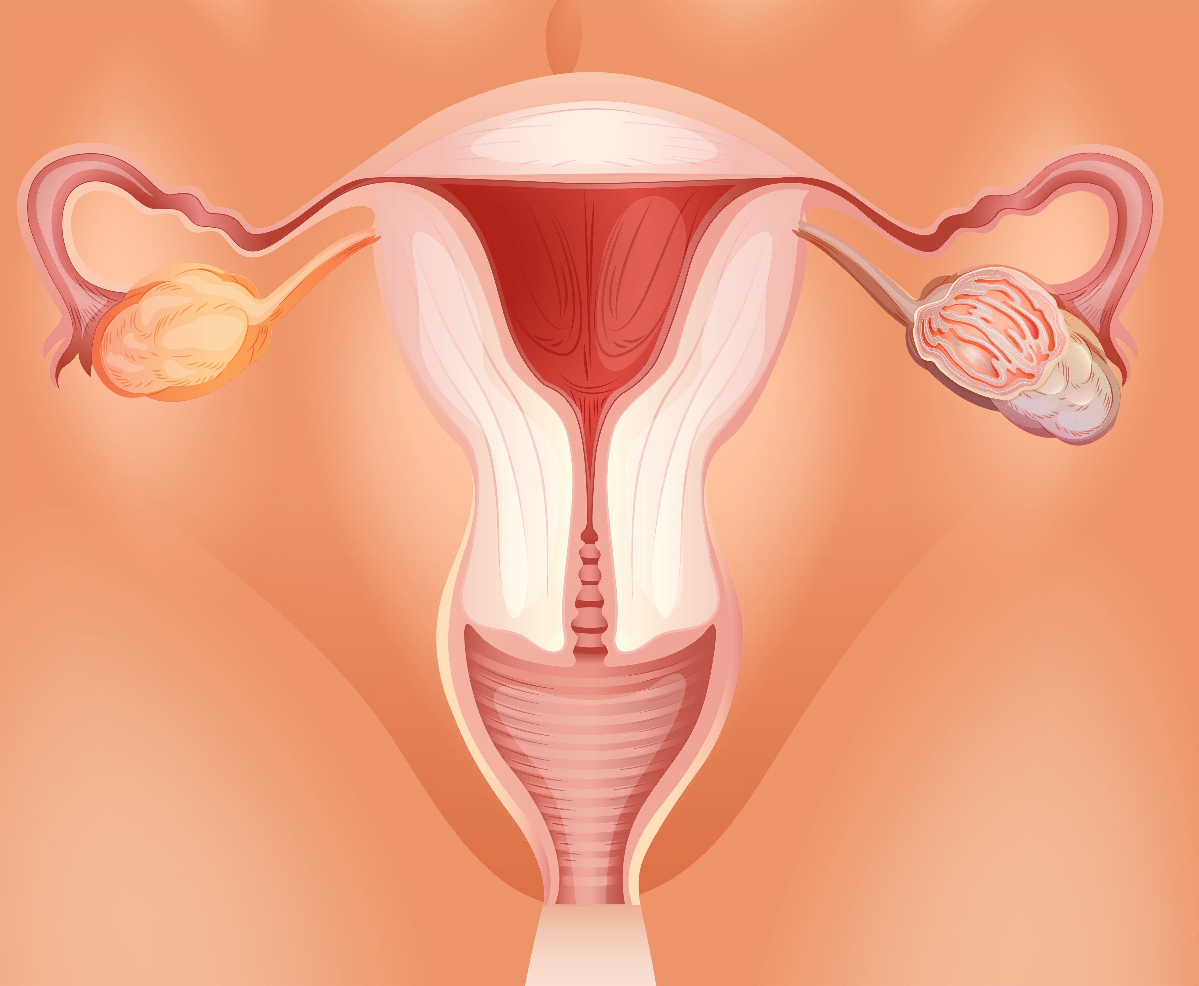
 Patient survival with ovarian tumors ranges from 30% to 50%.1 Therefore, there is a growing interest in patient-derived xenografts as a more accurate preclinical platform because of skepticism regarding the predictive capacity of xenografts derived from cell lines. This is because patient-derived xenografts are not only an accurate phenocopy of the patient’s tumor, but are also a copy at expression level, reflect the specific tumor mutations and preserve copy number variants for multiple generations. However, an ongoing challenge with patient-derived xenografts of tumors is their rate of engraftment, which can take anywhere from 1 month to 10. Furthermore, cancers such as ovarian tumors do not have equal representation of histological subtypes of patient populations, and pre-clinical testing requires large cohorts of histologically identical patients. Researchers therefore need reliable preservation methods for patient-derived xenografts of ovarian tumors. Typical patient-derived xenograft biobank samples use an fetal calf serum (FCS)/DMSO protocol, but the growth and take rate remains an unknown. Alkema et al. (2015)2 compare this to a vitrification-based protocol using 10% FCS and stepwise increasing concentrations of DMSO, propanediol, polyvinylpyrrolidone and ethylene glycol.
The investigators collected 66 advanced stage (III/IV) ovarian cancers and implanted them in mice. They had a take rate of 68% (n=45). The most notable predictive factors for successful engraftment was whether tissue was exposed to chemotherapy and the percentage of vital tumor cells. In 60% of those that did not engraft, the vital tumor cell percentage was less than 10%. Once engrafted tumors grew to 1 cm3, the investigators harvested them and re-transplanted into additional mice to establish further generations, where they showed successful engraftment in further generations. They selected eight ovarian tumors for an in-depth study, cut into 6 to 10 pieces, for storage using vitrification and/or FCS/DMSO. They transplanted 34 pieces from F1 patient-derived xenografts, representing all eight tumors, 91% of which grew. Using the vitrification method on F1 material, four out of five samples engrafted successfully; however, the overall take rate of implanted tumor pieces was 38% (8/21 tumor pieces). The FCS/DMSO method was more successful at the take stage. Two out of three tumors engrafted successfully and had a take rate of 67%. Alkema et al. then harvested the mouse tumors and stored them using the vitrification and/or the FCS/DMSO method, followed by re-engraftment of F2 generations of mice. All F2 generations were successful. However, they found better take rates using the FCS/DMSO protocol (94%) when compared to the vitrification (67%). For vitrified primary tumors, the latency time period of F1 generation varied from 70 to 320 days, whereas with FCS/DMSO it varied between 18 and 220 days.
Using immunohistochemistry, the researchers found that the proliferative rate was preserved through generations as well as in tissue engrafted after storage for both vitrification and FCS/DMSO. Furthermore, neither storage method influenced estrogen receptor and progesterone receptor expression. However, progesterone receptor expression increased in F2 generation compared to primary tumors.
A comparison of copy number alterations (CNAs) showed grafted tumors maintained the CNA pattern of the parental patient tumor; however, there was marked heterogeneity between patients. From this, Alkema et al. posit that ovarian cancer patient-derived xenografts of tumors retain their genomic characteristics during propagation over several generations.
References
1. World Cancer Research Fund International (2015) “Ovarian cancer statistics.”
2. Alkema et al. (2015) “Biobanking of patient and patient derived xenograft ovarian tumour tissue: Efficient preservation with low and high fetal calf serum based methods,” Science Reports, 5 (14495). doi: 10.1038/srep14495
Leave a Reply