Introduction
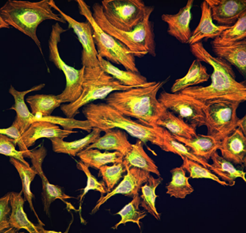
Cell painting, an innovative image-based profiling tool, was introduced in 2013 by Gustafsdottir S. M. et al and later by Anne Carpenters lab at the BROAD Institute in 2016. This assay is designed to facilitate the development of drug discovery assays and multiparameter testing campaigns by extracting a wealth of morphological and functional data from cells. The Invitrogen™ Image-iT™ Cell Painting Kit has revolutionized our approach to phenotypic surveillance, particularly when paired with the sophisticated Thermo Scientific™ CellInsight™ CX7 LZR Pro Platform.
Origins of the cell painting assay
Cell painting was first introduced in 2013 and later made popular by a 2016 Nature Protocols paper by Anne Carpenter’s lab. This assay can extract measurements from each cell based on changes in size, shape, texture, and fluorescence intensity. These measurements can detect subtle changes in phenotype, making cell painting a powerful tool for high-content analysis in drug discovery. The assay has undergone several iterations and continues to be a cornerstone technique.
Why the cell painting assay is useful for drug discovery
The cell painting assay’s utility lies in its ability to generate rich, multidimensional data from cell cultures. By using a combination of dyes to stain various cellular components, researchers can capture a comprehensive snapshot of cell morphology and organization. This is comprehensive view is especially helpful in drug discovery for several reasons including:
- Phenotypic profiling: Understanding the effects of genetic modifications or drugs on cell morphology can reveal potential insights into the clinical management of disease.
- High-throughput analysis: The assay enables the identification of hits in initial discovery screens by observing the impact of compounds on cell structures.
- Mechanistic insights: The data-rich output enables mechanistic insights into how drugs affect cellular components, which is often unavailable through traditional high-throughput assays.
The assay can generate information on over 1,500 features of the cell, enabling rapid data generation for the drug discovery pipeline. This capability makes it widely used in both pharmacological and academic research due to the sheer amount of data that can be generated quickly.
Challenges and overcoming them
Despite its advantages, the cell painting assay presents some challenges, primarily related to spectral overlap of the fluorescent dyes used. This overlap can hinder the accurate quantification of certain cellular targets. To address these issues, advancements in near-infrared reagents and imaging platforms, along with platform-specific reagent optimization, can significantly reduce spectral interference. The Thermo Scientific CellInsight CX7 LZR Pro Platform, with its advanced imaging capabilities, offers a solution to these challenges by helping provide higher resolution and more precise quantification of cellular components.
Case study: p53 knockout in A549 cells for drug discovery
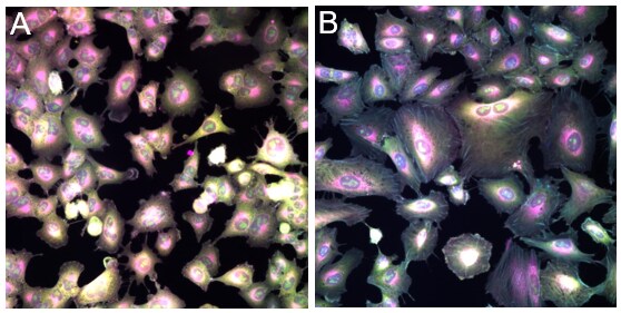
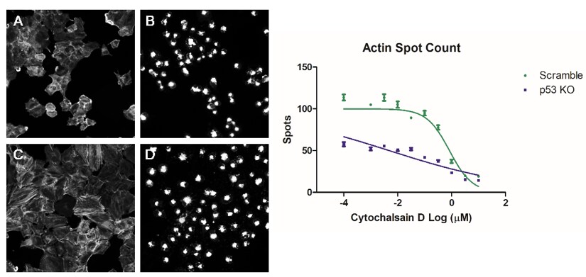
Using the optimized cell painting assay on the CellInsight CX7 LZR Pro, we can explore phenotypic differences between A549 wild-type cells and p53 knockout (KO) A549 cells. This comparison is crucial for understanding how the absence of functional p53 affects cellular responses to various drugs and compounds, which is essential in the context of drug discovery.
Observations
- Morphological changes: The absence of p53 can lead to noticeable changes in cell morphology, including alterations in cell size, shape, and internal organization.
- Drug sensitivity: p53 KO cells may exhibit different sensitivities to chemotherapeutic agents, helping provide insights for further research into therapeutics for p53-mutant cancers.
Key considerations for beginning cell painting assays in drug discovery
When starting with cell painting assays, several key elements should be considered:
- Reagent selection: The Invitrogen Image-iT Cell Painting Kit includes six reagents, but incorporating new dyes can deepen your analysis. Multiplexing abilities can be expanded with other labels and antibodies.
- Spectral overlap: Understanding the limitations of your imaging platform in discerning spectral overlap is crucial for accurate quantification.
- Protocol optimization: The existing protocol combines live and fixed cell staining steps. Streamlining this process, such as developing fixed cell mitochondrial stains, can enhance efficiency.
- Automated image analysis: Utilizing automated image analysis software to identify individual cells and measure morphological features helps ensure consistent and accurate data collection.
Assay outlook
The advancement of the cell painting assay in drug discovery looks promising, with potential advancements including:
- Enhanced imaging technologies: Continued improvements in imaging platforms can further reduce spectral overlap and enhance resolution.
- AI and machine learning: Integrating AI for data analysis can streamline the interpretation of complex phenotypic data, leading to more rapid and accurate conclusions.
- Expanded applications: The assay can be adapted for use in a wider range of cell types and conditions, broadening its applicability in biomedical research and drug discovery.
Software packages with improved automation and AI will further enhance the utility and importance of this assay. Optimization of dyes for specific platforms will also allow for greater accuracy and quantification by eliminating spectral overlap. Additionally, improvements in hardware and hyperspectral imaging capabilities will enable more targets and phenotypes to be evaluated simultaneously.
Conclusion
The Invitrogen Image-iT Cell Painting Kit, combined with the Thermo Scientific CellInsight CX7 LZR Pro Platform, represents a powerful tool for high-content testing in drug discovery. By overcoming current challenges and leveraging advanced technologies, researchers can gain deeper insights into cellular phenotypes and their implications for disease research. As we continue to refine and expand the capabilities of the cell painting assay, its role in advancing our understanding of cellular biology and drug discovery will undoubtedly grow.
References
- Thermo Fisher Scientific. (2022). Cell painting high-content screening assay: Application note. Retrieved from https://assets.thermofisher.com/TFS-Assets/BID/Application-Notes/cell-painting-high-content-screening-assay-app-note.pdf
- Bray, M. A., Singh, S., Han, H., et al. (2016). Cell painting, a high-content image-based assay for morphological profiling using multiplexed fluorescent dyes. Nature Protocols, 11(9), 1757–1774. https://doi.org/10.1038/nprot.2016.105
For Research Use Only. Not for use in diagnostic procedures.
© 2025 Thermo Fisher Scientific Inc. All rights reserved. All trademarks are the property of Thermo Fisher Scientific and its subsidiaries unless otherwise specified.

Thermo Fisher Scientific Partners with AIM Biotech on Development of Microphysiological Systems
The adoption of New Approach Methodologies (NAMs) in drug di...
Read More
5 Solutions for Overcoming Research Challenges in Vaccine Development
What are the main challenges in vaccine development? The mai...
Read More
Spatial Omics Meets Neuroscience: Dual ISH-IHC Brain Mapping Case Study
Spatial omics biology is reshaping how researchers study com... Dana D'Amico
Read More
APD vs PMT and Beyond: Flow Cytometry Light Detectors
Flow cytometry relies on sensitive photodetectors to convert... Aram Schiffman Eric Finan
Read More
Leave a Reply