In the world of materials science, micro computed tomography (microCT) uses X-rays to examine the inside structures of composite objects at high resolution. The process typically works because of the difference in absorption rates between two objects—say a piece of metal embedded in a matrix of polymers. Because the metal absorbs the x-rays at a higher rate than the surrounding material, researchers get the high-contrast images they need.
But what happens if there’s not a big difference in absorption rates? That’s exactly the challenge of microCT when it comes to fiber-reinforced polymers. Using microCT, industrial manufacturers in industries ranging from aerospace to automotive to energy test fiber-reinforced polymers to obtain insights into their microstructures. Yet because the fibers absorb the x-rays at around the same rate as the surrounding resin, it’s difficult to get the high contrast needed for a clear visualization.
Another challenge is the amount of energy required to obtain high-quality images for dense metals such as stainless steel without letting too many x-rays pass through the polymer and oversaturating the signal. This, in turn, generates a lot of background noise and distorts the resulting image.
The good news is that our HeliScan microCT for Materials Science solves these problems. The HeliScan provides advanced helical scanning and iterative reconstruction technology to produce unsurpassed image fidelity. The HeliScan system includes an x-ray source of between 20 and 160 kV, allowing users to tune down the energy and avoid oversaturating the signal. It also offers phase contrast retrieval, retrieving phase shift information—or data regarding changes in the phase of an x-ray beam that passes through an object—to help solve the low contrast problem.

Figure 1. Standard reconstruction (left) vs. phase contrast retrieval reconstruction (right) of a fiber-reinforced polymer.
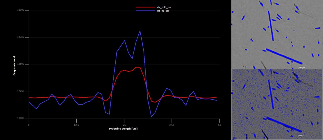
In the standard reconstruction in Figure 1, the signal to noise ratio does not allow for the automatic separation of the fibers from the resin matrix. By comparison, the phase contrast retrieval reconstruction accurately separates the two materials, and the background noise is automatically eliminated, leading to a far clearer and more accurate image.

Figure 2. The HeliScan automatically eliminates all information below the user’s designated threshold. In the above histogram, all data below the yellow line is removed, producing the clear image on the upper righthand side compared to the one below it in which the background noise has not been removed.
The HeliScan eliminates background noise by allowing users to establish their own threshold based on the greyscale result and then rejecting all the information beneath it. In Figure 2, all of the data below the yellow line is eliminated, automatically removing the background noise and generating the clearer image shown on the upper righthand side.
By solving the contrast problem and eliminating background noise, the HeliScan enables industrial manufacturers to automatically segment out composite materials in a matter of minutes compared to the hours required to do so manually. As a result, they can now examine fiber-reinforced polymers far more efficiently and precisely than was possible in the past—leading them to easily identify failures and ultimately design stronger, more durable materials.
To learn more about the high-fidelity microCT images generated by the HeliScan, fill out this form to speak with an expert.
Related articles:
- MicroCT: X-ray Vision at the Micro-Scale
- Building a Better Battery with MicroCT
- Improving the Additive Manufacturing Process with MicroCT
Dirk Laeveren is Product Marketing Manager, MicroCT at Thermo Fisher Scientific.
Subscribe now to receive Accelerating Microscopy updates straight to your inbox.
Leave a Reply