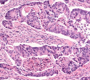
Tyrosine kinase fibroblast growth factor (FGF) receptors (FGFRs) are transmembrane proteins that bind to FGFs, leading to increased kinase activity and autophosphorylation of tyrosine residues. One epithelial variant of FGFR2, FGFR2b (or keratinocyte growth factor receptor), may be activated by both FGF-7 and FGF-10.1 Dysregulated FGFR2b has been linked to skeletal disorders and cancer.2,3
Recently, Francavilla et al. (2013) used functional assays and liquid chromatography–tandem mass spectrometry (LC-MS/MS) on LTQ Orbitrap and Q Exactive instrumentation (Thermo Scientific) to identify 971 phosphorylated tyrosine sites and evaluate the link between receptor trafficking and the fate of the cell.4
To do this, the researchers first transfected HeLa cells with FGFR2b and observed opposite signaling dynamics in FGF-7 and FGF-10. Stimulation with FGF-7 produced transient activation of FGFR signaling effectors, including sustained Erk phosphorylation, which is linked to cell proliferation. Stimulation with FGF-10 induced prolonged FGFR signaling without activation of FGFR1 or subsequent Erk phosphorylation, which is linked to cell migration.
The researchers then used stable isotope labeling by amino acids in cell culture (SILAC) and MS/MS to analyze tyrosine phosphoproteome changes after stimulation with FGF-7 and FGF-10 at 1 minute, 8 minute, and 40 minute intervals. The majority (84%) of the tyrosine residues were phosphorylated after 1 minute of stimulation. They identified 1,212 unique peptides with tyrosine phosphorylation and 971 phosphorylated tyrosine sites mapped to 624 proteins.
They analyzed this data set with principal component analysis (PCA) and fuzzy c-means clustering. They observed similar responses between FGF-7 and FGF-10 at each interval (1 min, 8 min, 40 min) and that 323 of the 971 sites possessed a ratio for each of the time points. They noted that 46% of these sites differentially clustered when stimulated with FGF-7 or FGF-10. Gene Ontology (GO) enrichment of each cluster revealed that FGF-10 stimulation induced events related to cell migration, branching and recycling, while FGF-7 stimulation induced events related to cell proliferation.
Francavilla et al. then transfected cells with wild-type and mutant FGFR2b in order to evaluate the role Y734 phosphorylation plays in FGFR2b signaling. They found that FGF-10 stimulation with mutant FGFR2b resulted in transient (rather than prolonged) signaling. Because signaling duration is tied to either receptor degradation or recycling,5 the researchers used immunofluorescent microscopy to observe that, after 90 minutes of stimulation with FGF-7, FGFR2b was not detectable in the cytoplasm or cell surface, which is consistent with receptor degradation.6 After the same amount of stimulation with FGF-10, FGFR2b recycled to the cell surface. Francavilla et al. state that these data indicate that FGF-10 stimulation induces Y734 phosphorylation that acts as a trigger for signaling duration and subsequent receptor sorting.
The researchers also used SILAC-labeled lysates and LC/MS to elucidate the recruitment of phosphoinositide 3-kinase (PI3K) and SH3 binding protein 4 (SH3BP4) to form a Y734-phosphorylated FGFR2b-PI3K-SH3BP4 complex following stimulation with FGF-10. They observed that FGF-10 stimulation resulted in formation of an FGFR2b-p85 (PI3K-recruiting peptide) complex, while FGF-7 stimulation did not. They confirmed this observation using breast cancer lines. Of 21 cell lines, 9 expressed varying levels of endogenous FGFR2b; 6 of these formed the FGFR2b complex upon stimulation by FGF-10 (but not with FGF-7). They also noted that mutant FGFR2b-Y734 (non-phosphorylated) failed to recruit p85.
Next, the team analyzed the roles that signaling duration and receptor sorting play in the opposite responses observed in FGF-7 and FGF-10 stimulation. They discerned that, in the presence of mutant Y734, FGF-10 stimulation produced less migration and increased proliferation, as compared to wild-type cells. They also observed that FGF-10 produced migration of breast cancer cells via FGFR2b-p85 complex interaction, while FGF-7 did not. They noted that prolonged decreases in SH3BP4 did not affect activities mediated by FGF-7 but, for FGF-10, did result in increased proliferation and reduction of migration.
Finally, the researchers examined epithelial branching in lung explants from embryonic mice. Treatment with FGF-7 resulted in cysts and bud expansion, while treatment with FGF-10 resulted in epithelial branching. When stimulated in the presence of the FGFR2b-Y734 mutant, treatment with FGF-10 resulted in bud expansion similar to treatment with FGF-7. Overall, the authors conclude that the FGFR2b-PI3K-SH3BP4 complex acts as a switch that governs receptor recycling or degradation.
Francavilla et al. assert that this research underlines the functional value of proteomics-based approaches for evaluating RTK signal transduction pathways. They also point to the therapeutic potential of ligand-mediated interactions such as those seen with FGFR2b and other signaling molecules. In particular, they present FGFR2b-Y734 phosphorylation and FGF-10 receptor adaptors as potential targets for future breast cancer research. Additional investigation into endocytotic regulators and receptor adaptors may also allow researchers and physicians to increase therapeutic efficiency and personalization.
References
1. Zhang, X., et al. (2006) “Receptor specificity of the fibroblast growth factor family: The complete mammalian FGF family,” Journal of Biological Chemistry, 281 (pp. 15694–700).
2. Katoh, M. (2008) “Cancer genomics and genetics of FGFR2 (Review),” International Journal of Oncology, 33 (pp. 233–7).
3. Wesche, J., Haglund, K., and Haugsten, E.M. (2011) “Fibroblast growth factors and their receptors in cancer,” Biochemical Journal, 437 (pp. 199–213).
4. Francavilla, C., et al. (2013) “Functional proteomics defines the molecular switch underlying FGF receptor trafficking and cellular outputs,” Molecular Cell, 51 (pp. 707–22).
5. Sigismund, S., et al. (2012) “Endocytosis and signaling: cell logistics shape the eukaryotic cell plan,” Physiological Reviews, 92 (pp. 273–366).
6. Belleudi, F., et al. (2007) “Keratinocyte growth factor receptor ligands target the receptor to different intracellular pathways,” Traffic, 8 (pp. 1854–72).
Post Author: Melissa J. Mayer. Melissa is a freelance writer who specializes in science journalism. She possesses passion for and experience in the fields of proteomics, cellular/molecular biology, microbiology, biochemistry, and immunology. Melissa is also bilingual (Spanish) and holds a teaching certificate with a biology endorsement.
New Diagnostic Biomarkers in Colorectal Cancer
Colonoscopies are currently the best method to diagnose colo...
Read More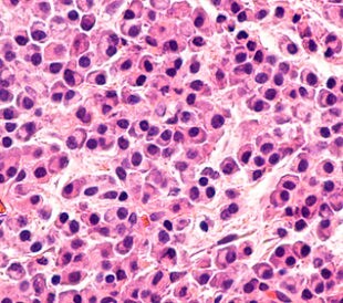
Top-Down Proteomic Characterization of Histone H3 Proteoforms in Disease
Zheng et al. performed top-down mass spectrometric proteomic...
Read More
Effects of Rosemary Extract on Colon Cancer Cells
A team of researchers from Spain and Sweden recently ...
Read More
Lung Cancer Biomarker Discovery through Metabolic Enzyme Activity
Sun et al. (2016) present a biomarker discovery study that s...
Read More
Leave a Reply