Chaperone proteins are responsible for proper protein folding and have been well studied in Saccharomyces cerevisiae. To date, 63 proteins have been characterized and 4,340 candidate substrates have been identified.1 However, the qualitative map of the yeast proteome lacks quantification.
In their recent publication, Brownridge et al.2 were able to quantify 51 of the 63 annotated chaperones in the yeast proteome over a range of 250 to 440,000 copies per cell. Quantification included coverage over all chaperone classes, including proteins that previous studies have failed to quantify, such as HSP70 protein Ssa3 and CCT proteins. They also were able to quantify three small class chaperones that previously have remained unreported.
For their quantification studies, Brownridge et al.2 developed proteins that contained concatenating protein sequences of target proteins added to artificial proteins (QconCAT). QconCAT proteins were able to act as an internal standard to enable quantification of several proteins at a time.3
Cultured Saccharomyces cerevisiae was digested, and Top3′ label-free analysis was performed on two platforms: an ion-mobility-coupled data-independent (HDMSE) method on Synapt G2 and a data-dependent method on a Q-Exactive (Thermo Scientific).
Three recombinant proteins were designed to quantify chaperone proteins. Heavy QconCAT proteins containing stable-isotope-labeled amino acids were generated and spiked into yeast protein samples to enable quantification. These samples were analyzed using selected reaction monitoring (SRM) on a nano Acquity UPLC system (Waters) coupled to a Xevo TQ triple quadrupole mass spectrometer (Waters).
The dilution was prepared by serial dilution of the yeast-QconCAT codigest by the unspiked yeast digest. Samples containing the protein equivalent of 200,000 cells with either 0.2, 2, or 20 fmol of QconCAT were analyzed by SRM. Spiked yeast digests were analyzed by LC/MS on an Ultimate 3000 RSLC nano system (Thermo Scientific) coupled to a Q-Exactive mass spectrometer (Thermo Scientific)
Values obtained from the label-mediated QconCAT approach were compared to other MS-based studies and epitope-tagging approaches. Though not able to quantify as many proteins as the QconCAT approach, the SILAC two-label-mediated approach employed by de Godoy et al.4 verified the data and served as a reference for obtained quantification values.
Through this work, Brownridge et al.2 were able to identify a correlation between chaperone protein abundance and the workload each chaperone performs. This information has importance in understanding the interactome of biological systems.
References
1. Gong, Y., et al. (2009) ‘An atlas of chaperone-protein interactions in Saccharomyces cerevisiae: implications to protein folding pathways in the cell‘, Molecular Systems Biology, 5 (275), doi/10.1038/msb.2009.26.
2. Brownridge, P., et al. (2013) ‘Quantitative analysis of chaperone network throughput in budding yeast‘, Proteomics, published online February 19, 2013. doi: /10.1002/pmic.201200412
3. Silva, J.C. (2005) ‘Quantitative proteomic analysis by accurate mass retention time pairs‘, Analytical Chemistry, 77 (7) (pp. 2187-2200)
4. de Godoy, L.M., et al. (2008) ‘Comprehensive mass-spectrometry-based proteome quantification of haploid versus diploid yeast‘, Nature, 455 (7217), (pp. 1251-1254)
Analysis of Ketosteroids by Girard P Derivatization
Dehydroepiandrosterone (DHEA); 4-androstene-3,17-dione (AD);...
Read More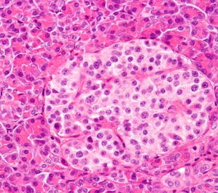
Profiling Human Islet Cells with Laser Capture Microdissection
Composed mainly of β-cells, human islet cells secrete h...
Read More
Intact Proteins in Native Conditions with Quadrupole-Orbitrap Mass Spectrometry
Successful research focusing on antibody and drug interactio...
Read More
Autophagy Helps Control Endothelial Permeability
A research team in Glasgow has identified autophagy as a str...
Read More
Leave a Reply