Search
Invitrogen
Phospho-S6 (Ser235, Ser236) Monoclonal Antibody (cupk43k), PE-Cyanine7, eBioscience™
{{$productOrderCtrl.translations['antibody.pdp.commerceCard.promotion.promotions']}}
{{$productOrderCtrl.translations['antibody.pdp.commerceCard.promotion.promocode']}}: {{promo.promoCode}} {{promo.promoTitle}} {{promo.promoDescription}}. {{$productOrderCtrl.translations['antibody.pdp.commerceCard.promotion.learnmore']}}
{{$ctrl.translations['antibody.pdp.skuNotes.additionalInformation'] || 'Additional Information'}}:
{{$ctrl.translations['antibody.pdp.skuNotes.additionalInformation']}}:

FIGURE: 1 / 2
Phospho-S6 (Ser235, Ser236) Antibody (25-9007-41) in Flow
Intracellular staining of freshly-harvested (left), unstimulated (middle), or 30-minute LPS-stimulated (right) normal human peripheral blood cells with Anti-Human CD14 eFluor® 450 (Product # 48-0149-42) and Anti-Human/Mouse phospho-S6 Ribosomal (S235/S236) PE-Cyanine7, using the Intracellular Fixation and Permeabilization Buffer Set (Product # 88-8824-00) and protocol. Cells in the lymphocyte/ monocyte gate... View More


Please note: We are reviewing Western blot images included in the antibody testing data in our catalog, including those provided by third parties. Unless expressly labeled or annotated as “raw-unedited”, Western blot images included in the antibody testing data in our catalog may have been edited, optimized or otherwise adjusted for presentation.


Product Details
25-9007-41
Applications
Tested Dilution
Publications
Product Specifications
Species Reactivity
Human,
Mouse
Published species
Human,
Mouse
Host/Isotype
Mouse
/ IgG1, kappa
Recommended Isotype Control
Class
Monoclonal
Type
Antibody
Clone
cupk43k
Conjugate
PE-Cyanine7
PE-Cyanine7
PE-Cyanine7
Excitation/Emission Max
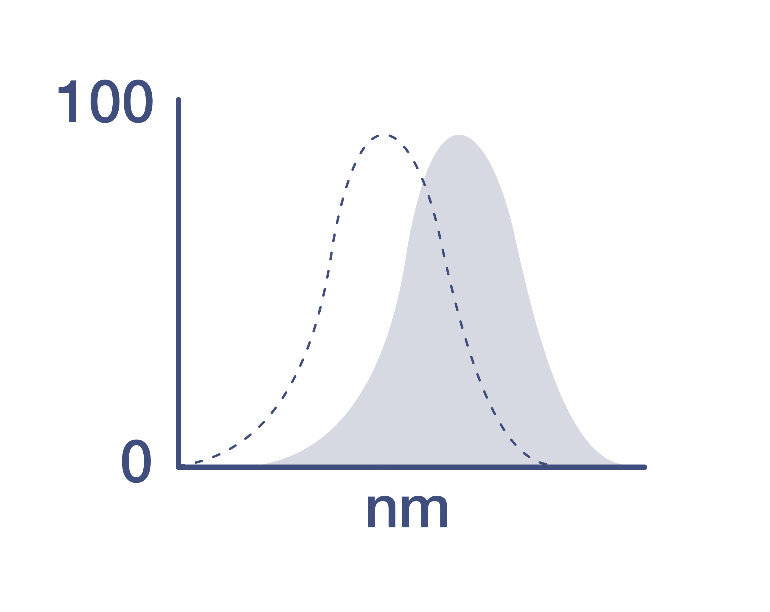
569/780 nm
View spectra
Form
Liquid
Concentration
5 µL/Test
Purification
Affinity chromatography
Storage buffer
PBS, pH 7.2, with BSA
Contains
0.09% sodium azide
Storage conditions
4°C, store in dark, DO NOT FREEZE!
Shipping conditions
Ambient (domestic); Wet ice (international)
RRID
Product Specific Information
Description: This cupk43k monoclonal antibody recognizes human and mouse ribosomal protein S6 (also known as 40S ribosomal protein S6, phosphoprotein NP33, RPS6, RS6, S6) when phosphorylated on serine 235 (S235, human) and serine 236 (S236, mouse). Ribosomal protein S6 is a component of the 40S subunit of the ribosome and is phosphorylated at multiple sites following stimulation of cells by growth factors, tumor promoting agents, or mitogens. Phosphorylation of ribosomal protein S6 by p70S6K and PKDCD results in upregulation of the translation of RNA coding for proteins involved in cell cycle entry. Ribosomal protein S6 is dephosphorylated upon growth arrest.
The specificity of the cupk43k monoclonal antibody was determined by western blotting.
Applications Reported: This cupk43k antibody has been reported for use in intracellular staining followed by flow cytometric analysis.
Applications Tested: This cupk43k antibody has been pre-titrated and tested by intracellular staining and flow cytometric analysis of stimulated normal peripheral blood cells. This can be used at 5 µL (0.125 µg) per test. A test is defined as the amount (µg) of antibody that will stain a cell sample in a final volume of 100 µL. Cell number should be determined empirically but can range from 10^5 to 10^8 cells/test.
Use of Protocol A: Two-step protocol: intracellular (cytoplasmic) proteins allows for the greatest flexibility for detection of surface and intracellular (cytoplasmic) proteins. Use of Protocol B: One-step protocol: intracellular (nuclear) proteins is recommended for staining of transcription factors in conjunction with surface and phosphorylated intracellular (cytoplasmic) proteins. Protocol C: Two-step protocol: Fixation/Methanol allows for the greatest discrimination of phospho-specific signaling between unstimulated and stimulated samples, but with limitations on the ability to stain specific surface proteins (refer to "Clone Performance Following Fixation/Permeabilization"). All Protocols can be found in the "Staining Intracellular Antigens for Flow Cytometry Protocol" located in the BestProtocols® Section under the Resources tab online.
Light sensitivity: This tandem dye is sensitive to photo-induced oxidation. Please protect this vial and stained samples from light.
Fixation: Samples can be stored in IC Fixation Buffer (Product # 00-8222) (100 µL of cell sample + 100 µL of IC Fixation Buffer) or 1-step Fix/Lyse Solution (Product # 00-5333) for up to 3 days in the dark at 4°C with minimal impact on brightness and FRET efficiency/compensation. Some generalizations regarding fluorophore performance after fixation can be made, but clone specific performance should be determined empirically.
Excitation: 488-561 nm; Emission: 775 nm; Laser: Blue Laser, Green Laser, Yellow-Green Laser.
Filtration: 0.2 µm post-manufacturing filtered.
Target Information
Ribosomes, the organelles that catalyze protein synthesis, consist of a small 40S subunit and a large 60S subunit. Together these subunits are composed of 4 RNA species and approximately 80 structurally distinct proteins. This gene encodes a cytoplasmic ribosomal protein that is a component of the 40S subunit. The protein belongs to the S6E family of ribosomal proteins. It is the major substrate of protein kinases in the ribosome, with subsets of five C-terminal serine residues phosphorylated by different protein kinases. Phosphorylation is induced by a wide range of stimuli, including growth factors, tumor-promoting agents, and mitogens. Dephosphorylation occurs at growth arrest. The protein may contribute to the control of cell growth and proliferation through the selective translation of particular classes of mRNA. As is typical for genes encoding ribosomal proteins, there are multiple processed pseudogenes of this gene dispersed through the genome.
For Research Use Only. Not for use in diagnostic procedures. Not for resale without express authorization.
How to use the Panel Builder
Watch the video to learn how to use the Invitrogen Flow Cytometry Panel Builder to build your next flow cytometry panel in 5 easy steps.
Bioinformatics
Protein Aliases: 40S ribosomal protein S6; OTTHUMP00000021121; OTTHUMP00000021122; phosphoprotein NP33; ribosomal protein S6; RP11-513M16.6; RS6; unnamed protein product
Gene Aliases: eS6; S6; S6R
UniProt ID: (Human) P62753, (Mouse) P62754
Entrez Gene ID: (Human) 6194, (Mouse) 20104
ribosomal small subunit assembly
G1/S transition of mitotic cell cycle
mitotic cell cycle
placenta development
cytoplasmic translation
T cell proliferation involved in immune response
rRNA processing
translation
activation-induced cell death of T cells
gastrulation
positive regulation of cell proliferation
oogenesis stage
TOR signaling
response to insulin
T cell differentiation in thymus
ribosome biogenesis
ribosomal small subunit biogenesis
glucose homeostasis
positive regulation of apoptotic process
negative regulation of apoptotic process
erythrocyte development
cellular response to ethanol
negative regulation of bicellular tight junction assembly
Disclaimer

Clicking the images or links will redirect you to a website hosted by BenchSci that provides third-party scientific content. Neither the content nor the BenchSci technology and processes for selection have been evaluated by us; we are providing them as-is and without warranty of any kind, including for use or application of the Thermo Fisher Scientific products presented.

Performance Guarantee
If an Invitrogen™ antibody doesn't perform as described on our website or datasheet,we'll replace the product at no cost to you, or provide you with a credit for a future purchase.*
Learn more
We're here to help
Get expert recommendations for common problems or connect directly with an on staff expert for technical assistance related to applications, equipment and general product use.
Contact tech support