Search


Citations & References (32)
Invitrogen™
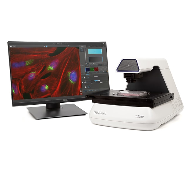
EVOS™ M7000 Imaging System
The EVOS M7000 Imaging System is a high-performance, fully automated, inverted, multi-channel fluorescence and transmitted light imaging system. It wasRead more
| Catalog Number | Quantity |
|---|---|
| AMF7000 | 1 system |
Catalog number AMF7000
Price (HKD)
-
Quantity:
1 system
The EVOS M7000 Imaging System is a high-performance, fully automated, inverted, multi-channel fluorescence and transmitted light imaging system. It was developed to meet today's expectations for image quality, user interface interactions, data generation speed and flexibility, and end-user value. The powerful and time-saving acquisition tools with enhanced autofocus algorithms and automated routines for microwell plate assays, time-lapse live cell imaging, area scanning in time lapse and Z-stacks in scan mode, and a superb optomechanical design enable the generation of publication-quality images and data with just a few clicks when and where needed.
The EVOS M7000 Imaging System offers these advantages:
- Outstanding image quality and versatility with a 5-position objective turret, 4-color LED fluorescence and transmitted light channels, and 3.2 MP CMOS color and B/W cameras
- Exceptional usability with fully automated X/Y scanning stage, autofocus, and acquisition routines
- High-speed image acquisition coupled with multi-position well scanning, and Z-stack and tile-stitch options lend power and flexibility to your data generation
- Fully integrated time-lapse live cell imaging using the optional EVOS Onstage Incubator for precise control of temperature, gases for normoxic or hypoxic conditions, and humidity
- Powerful image analysis capabilities for cell segmentation and quantification with the optional Celleste Image Analysis Software
- EVOS Analysis application allows for annotation, cell count, and 2D deconvolution of images and is provided as standard
Fully automated imaging system
The EVOS M7000 system combines and integrates precision components with a unique modern design functionality that enables high-quality automated fluorescence imaging anywhere with unprecedented workflow efficiency. Full automation of the precision X/Y-stage movement; changing of the 4 LED fluorescent light cubes, 5-position objective turret, focus, and exposure; and switching of the dual camera make the EVOS M7000 system an exceptional platform for a variety of demanding imaging applications. Through the intuitive acquisition interface, you can program the EVOS M7000 system to run well-plate scanning, time lapse experiments, and tile-stitch/montage area scans in Z-stack and/or time-lapse modes in your vessel of choice. For live cell imaging over multiple hours or days, the optional EVOS Onstage Incubator, which functions as an environmental chamber on top of the microscope stage, is operated seamlessly from within the instrument interface. The EVOS M7000 system is compatible with all accessories for the predecessor EVOS FL Auto imaging system.
Versatile and highly configurable
With a 5-position objective turret and the ability to simultaneous acquire images in four fluorescence channels plus transmitted light, the optical system can be easily configured to meet your needs. Choose from our broad range of high quality coverslip-corrected and long-working-distance objectives for use with plastic vessels or glass sides. Available from 1.25X to 100X magnification in achromat, fluorite, or apochromat models, together with over twenty LED light cubes, the sample imaging options are limitless. Dual monochrome and color high-sensitivity dual CMOS cameras enable seamless imaging of both fluorescent and chromogenically stained samples. Interchangeable vessel holders accommodate most vessel types and sizes, including slides, multi-well plates, culture flasks, and Petri dishes, and afford precise control and sample alignment by the automated stage. The vessel creation wizard in the user interface enables creation of profiles for custom sample vessels.
Powerful software
The EVOS M7000 software presents users with an extensive suite of tools for acquisition, visualization, and analysis. The user interface was designed with a strong emphasis on usability, including an option to view samples in a single field mode (Field View) or in a window that covers a much larger area (Area View). By using a zoom function, the Area View size can be adjusted. A user can explore a large sample area at low magnification, define regions of interest, and then seamlessly transition to the Scan function in Automate Mode and execute an area scan at higher magnification.
The enhanced power of the EVOS M7000 system enables more demanding automated acquisition routines like area scanning in time lapse and Z-stacks in scan mode to be performed. Visualization capabilities include multi-field image viewing gallery, mosaic tiling and tile stitching options, Z-stack visualization, and a video recording and movie maker/editor for time lapse sequences. The EVOS M7000 system is fully compatible with the EVOS Onstage Incubator environmental chamber for control of temperature, gases, and humidity. With the ability generate both hypoxic and normoxic conditions, it is possible to study live cells over extended periods with highly flexible time lapse automation routines.
For users needing more powerful image analysis capabilities, images acquired on the EVOS M7000 system can be analyzed directly using the optional Celleste Image Analysis Software. Celleste software offers powerful tools for image segmentation and classification that can be applied to a range of cell analysis applications for counting and measuring intensity, area, and shape changes over time, and more.
Smart LED illumination technology
All EVOS fluorescence imaging systems utilize our proprietary LED light cubes. This light engine outputs remarkable intensity over a short light path and delivers incomparable fluorophore excitation. Each light cube contains a precisely matched set of optical components to optimize the position, evenness, and intensity of the light beam. Digitally controlled LED light sources allow adjustment of illumination levels to the sample and experimental conditions to minimize the risk of photo-toxicity and photo-bleaching in time-lapse studies. Hard-coated filters give sharper edges and significantly higher transmission efficiencies than traditional soft-coated filters.
Easy to use and reliable
The EVOS M7000 Imaging System is powerful, yet offers the simplicity of plug-and-play. Like other EVOS systems, it requires no warm-up or cool-down periods. The LED light source guarantees exceptional stability and durability, so you can turn the unit on and off whenever you need to image a sample. The environmentally safe, mercury-free LED bulbs are rated for >50,000 hours, compared to 300 hours for a typical mercury bulb and 1,500 hours for a metal halide bulb. The long lifetime and low energy consumption translate into significantly lower operating costs compared to instruments with conventional light sources. The EVOS M7000 system is powered by an external Dell XE4 computer with Intel Core i9 processor, 128 GB DDR4 RAM, 4 TB SSD, and NVIDIA Quadro RTXA4000 with 8 GB discrete video graphics running Windows 10. Whether acquiring single images or setting up automated routines, the EVOS M7000 system is remarkably easy to use and run.
The EVOS imaging systems are built from the ground up to maximize performance and optimize workflow. You will be astonished at how easy it is to operate and amazed at how extraordinary your images look on-screen.
Explore the entire EVOS line of imaging systems, accessories, and offers ›
System and hardware specifications
- Optics:infinity‐corrected optical system; RMS‐threaded objectives with 45-mm parfocal distance
- Imaging mode: fluorescence, brightfield, color brightfield, and phase contrast
- Illumination: five position chamber for 4 fluorescence light cubes plus brightfield; light cubes with integrated hard coated filter set and LED light source with >50,000 hour life; broad selection of standard and specialty light cubes
- Imaging methods: single color, multi-color, area scan with montage or tile-stitch, time lapse, Z-stacking, movie capture
- Objective capacity: 5‐position turret
- Objectives (not included): wide selection of high‐quality LWD and coverslip‐corrected objectives
- Condenser:60-mm long working distance condenser, 4 position turret with a clear aperture and 3 phase annuli
- Stage: motorized X/Y scanning stage; travel range 120 mm x 80 mm with sub‐micron resolution, drop‐in inserts to receive vessel holders and lock down holders to fix sample in place during long scans
- Focus mechanism:automated focus mechanism with sub‐micron resolution
- LCD display: 27-inch 4K color monitor; 3840 x 2160 pixel resolution
- Cameras: high-sensitivity 3.2 MP (2048 x 1536) monochrome CMOS sensor with 3.45 μm pixel resolution;high-sensitivity 3.2 MP (2048 x 1536)
For Research Use Only. Not for use in diagnostic procedures.
Specifications
DisplayLCD
For Use With (Application)Neurobiology, Immuno-Oncology, Live-Cell Imaging, 3D Cell Imaging, High-Resolution Tile Scanning, Immunohistochemistry (IHC)
Light SourceLED
Magnification1.25X to 100X
ModelM7000
Objectives5 Position Turret
Quantity1 system
Resolution1920 x 1080 pixels
Shipping ConditionRoom Temperature
Camera3.2 MP Color CMOS
Product LineEVOS
TypeCell Imaging System
Unit SizeEach
Contents & Storage
EVOS M7000 Imaging System
- 27-inch 4K LCD monitor with 3840 x 2160 pixel resolution
- External Dell XE4 computer with Intel Core i9 processor, 128 GB DDR4 RAM, 4 TB SSD, and NVIDIA Quadro RTXA4000 with 8 GB discrete video graphics running Windows 10
EVOS M7000 Accessory Kit
- EVOS M7000 Quick Start Guide
- 16 GB USB 3.0 drive with user manual
- Universal power supply, 24V, 5A
- 3 North America (Type B) power cords*
- USB 3.0 type A to B cable
- Display port to display port cable, 6 ft
- Cube access tool
- EVOS calibration slide (AMEP4720)
- Vessel holder with retention for two slides (AMEPVH021)
- Vessel holder with retention for microwell plates (AMEPVH022). Also functions as adapter for vessel holders AMEPVH001 through AMEPVH018.
- Block condenser slider (AMEP4688)
- Diffuser condenser slider for brightfield applications (AMEPDFS1)
- Slider for 4X objectives (AMEP4738)
- EVOS light shield
- Dust cover
- Shipping restraint removal guide
- Mouse pad
- Tech Support card
- Corded optical USB mouse
- Keyboard
* Note that three (3) country-specific power cord must be ordered separately in regions not using the Type B (North America) power plug:
- AMEP4644 EVOS Power Cord, Type A (North America)
- AMEP4645 EVOS Power Cord, Type G (United Kingdom)
- AMEP4646 EVOS Power Cord, Type C (Europe)
- AMEP4647 EVOS Power Cord, Type I (Australia)
- AMEP4648 EVOS Power Cord, Type L (Italy)
- AMEP4649 EVOS Power Cord, Type H (Israel)
- AMEP4708 EVOS Power Cord, Type D (India/South Africa)
- AMEP4782 Power Cord, Type I (China, CCC certified)
- AMEP4786 Power Cord Type J (Switzerland)
Frequently asked questions (FAQs)
Can I visualize Alexa Fluor 488 dye using the EVOS M7000 Imaging System?
Can I upgrade my EVOS M5000 Imaging System (Cat. No. AMF5000)?
How do I turn on the 'phase mode' on the EVOS M7000 Imaging System for use with my phase objectives?
What warranty does the EVOS M7000 Imaging System come with?
During a scan routine with the EVOS M7000 Imaging System, I noticed that my fluorescent samples ended up out of focus. How can I correct this?
Citations & References (32)
Citations & References
Abstract
Punicalagin Prevents Inflammation in LPS-Induced RAW264.7 Macrophages by Inhibiting FoxO3a/Autophagy Signaling Pathway.
Journal:Nutrients
PubMed ID:31731808
'Punicalagin, a hydrolysable tannin of pomegranate juice, exhibits multiple biological effects, including inhibiting production of pro-inflammatory cytokines in macrophages. Autophagy, an intracellular self-digestion process, has been recently shown to regulate inflammatory responses. In this study, we investigated the anti-inflammatory potential of punicalagin in lipopolysaccharide (LPS) induced RAW264.7 macrophages and uncovered
In vitro assay for the efficacy assessment of AAV vectors expressing microdystrophin.
Journal:Exp Cell Res
PubMed ID:32360435
'AAV-delivered microdystrophin genes hold great promise for Duchenne muscular dystrophy (DMD) treatment. It is anticipated that the optimization of engineered dystrophin genes will be required to increase the efficacy and reduce the immunogenicity of transgenic proteins. An in vitro system is required for the efficacy testing of genetically engineered dystrophin
Co-regulation of the transcription controlling ATF2 phosphoswitch by JNK and p38.
Journal:Nat Commun
PubMed ID:33188182
'Transcription factor phosphorylation at specific sites often activates gene expression, but how environmental cues quantitatively control transcription is not well-understood. Activating protein 1 transcription factors are phosphorylated by mitogen-activated protein kinases (MAPK) in their transactivation domains (TAD) at so-called phosphoswitches, which are a hallmark in response to growth factors, cytokines
Escape from neutralizing antibodies by SARS-CoV-2 spike protein variants.
Journal:Elife
PubMed ID:33112236
'Neutralizing antibodies elicited by prior infection or vaccination are likely to be key for future protection of individuals and populations against SARS-CoV-2. Moreover, passively administered antibodies are among the most promising therapeutic and prophylactic anti-SARS-CoV-2 agents. However, the degree to which SARS-CoV-2 will adapt to evade neutralizing antibodies is unclear.
Clinical utility of circulating tumor-associated cells to predict and monitor chemo-response in solid tumors.
Journal:Cancer Chemother Pharmacol
PubMed ID:33170321
'Selection of cytotoxic chemotherapy agents (CCA) based on pre-treatment evaluation of drug sensitivities is a desirable but unmet goal for personalized anticancer treatment strategies. Prior attempts to correlate in vitro Chemo-Response Profiles (CRP) of tumor explants or Circulating Tumor Cells (CTCs) with clinical outcomes have been largely unsuccessful. We present