Search


Citas y referencias (8)
Invitrogen™
Click-iT™ Plus TUNEL Assay Kits for In Situ Apoptosis Detection
Detecte apoptosis en muestras de células y tejidos con kits de ensayo Click-iT Plus TUNEL, que ofrecen una fácil incorporación de colorantes y se pueden multiplexar con GFP y RFP.
Have Questions?
Cambiar vista

| Número de catálogo | Color | Etiqueta o tinte |
|---|---|---|
| C10619 | Rojo lejano | Alexa Fluor™ 647 |
| C10617 | Verde | Alexa Fluor™ 488 |
| C10618 | Rojo | Alexa Fluor™ 594 |
Número de catálogo C10619
Precio (MXN)
21,165.30
Each
Color:
Rojo lejano
Etiqueta o tinte:
Alexa Fluor™ 647
Precio (MXN)
21,165.30
Each
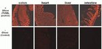
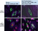
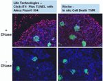
Detecte más células apoptóticas en tejidos y muestras de células cultivadas con el ensayo Click-iT Plus TUNEL para la detección de apoptosis in situ, que ofrece opciones de colorante fluorescente Alexa Fluor 488, 594 y 647. Este kit de detección de apoptosis in situ está optimizado para muestras de tejido o células y los colorantes se pueden multiplexar con otros colorantes o proteínas, como GFP y RFP, e incorporar más fácilmente en moléculas complejas debido a su tamaño más pequeño (en comparación con anticuerpos). Este kit de ensayo TUNEL también es muy flexible y se puede utilizar para probar de una a 50 muestras en un solo experimento.
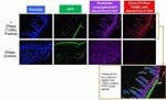
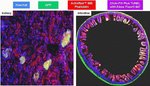
Los ensayos Click-iT Plus TUNEL Alexa Fluor 488, 594 y 647 para la detección de apoptosis in situ pueden detectar células apoptóticas en muestras de tejidos y células cultivadas mediante la incorporación de una pequeña fracción de etiquetado altamente específica y un colorante fluorescente brillante. Tras la incorporación de la fracción de etiquetado en los fragmentos de ADN, la detección se logra mediante una reacción de «clic» catalizada utilizando condiciones lo suficientemente leves como para preservar la señal fluorescente emitida de la GFP (proteína verde fluorescente) o RFP (proteína roja fluorescente).
Otras ventajas del ensayo Click-iT Plus TUNEL para detección de apoptosis in situ incluyen:
• Optimizado para la detección de células apoptóticas en muestras de tejido o células.
• El multiplexing permite trabajar de manera óptima con colorantes fluorescentes o proteínas como la GFP y RFP.
• Ensayo TUNEL mejorado: mejor incorporación de etiquetas debido a una pequeña fracción reactiva.
• Señal apoptótica brillante: usa coloraciones Alexa Fluor, las cuales provocan una señal fluorescente estable y sin decoloración fotográfica.
• Flexibilidad: el ensayo se puede configurar para probar de una a 50 muestras a la vez.
La fragmentación del ADN celular es un sello distintivo de la apoptosis. El ensayo TUNEL es el método más utilizado para detectar ADN fragmentado en células apoptóticas o muestras de tejido. El ensayo TUNEL comienza con la incorporación de dUTP (2'-desoxiuridina 5'-trifosfato) modificado en el extremo 3'-OH del ADN fragmentado. La modificación de dUTP suele ser la adición de un fluoróforo. Debido al tamaño del fluoróforo, la dUTP modificada puede mostrar unas tasas de incorporación inferiores a las esperadas, lo que puede afectar a la sensibilidad del ensayo TUNEL. Además, muchos de los fluoróforos utilizados en kits de ensayo TUNEL disponibles actualmente sufren de decoloración fotográfica y problemas de solapación espectral fluorescente, que reducen la sensibilidad y la capacidad de multiplex del ensayo.
El ensayo Click-iT Plus TUNEL ha sido desarrollado para dar respuesta a estas cuestiones. El ensayo utiliza dUTP modificada con un grupo alquino (un pequeño grupo funcional bioortogonal) que permite que el nucleótido se incorpore con más facilidad. Tras su incorporación, una reacción de clic muy específica entre el grupo alquino y un colorante fluorescente de azida picolil Alexa Fluor y la detección posterior de ese colorante, se obtiene como resultado un ensayo sensible y específico para la detección de células apoptóticas o muestras de tejido. A causa de sus condiciones de reacción suaves, el ensayo Click-iT Plus TUNEL permite el multiplexing con colorantes o proteínas fluorescentes.
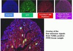
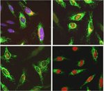
El ensayo Click-iT Plus TUNEL ha sido validado con varios tipos de tejidos fijados en formol incluidos en parafina. En todos los casos se conservó su capacidad de multiplexar con colorantes y proteínas fluorescentes. Además, se conservó la habilidad para la tinción de actina mediante faloidina etiquetada fluorescente.
El ensayo Click-iT Plus TUNEL contiene todos los reactivos necesarios para detectar células apoptóticas desde cualquier muestra de tejido o células. Los reactivos suministrados en este kit se pueden usar para probar 50 muestras y se pueden configurar para probar de una a 50 muestras a la vez.
Otras ventajas del ensayo Click-iT Plus TUNEL para detección de apoptosis in situ incluyen:
• Optimizado para la detección de células apoptóticas en muestras de tejido o células.
• El multiplexing permite trabajar de manera óptima con colorantes fluorescentes o proteínas como la GFP y RFP.
• Ensayo TUNEL mejorado: mejor incorporación de etiquetas debido a una pequeña fracción reactiva.
• Señal apoptótica brillante: usa coloraciones Alexa Fluor, las cuales provocan una señal fluorescente estable y sin decoloración fotográfica.
• Flexibilidad: el ensayo se puede configurar para probar de una a 50 muestras a la vez.
La fragmentación del ADN celular es un sello distintivo de la apoptosis. El ensayo TUNEL es el método más utilizado para detectar ADN fragmentado en células apoptóticas o muestras de tejido. El ensayo TUNEL comienza con la incorporación de dUTP (2'-desoxiuridina 5'-trifosfato) modificado en el extremo 3'-OH del ADN fragmentado. La modificación de dUTP suele ser la adición de un fluoróforo. Debido al tamaño del fluoróforo, la dUTP modificada puede mostrar unas tasas de incorporación inferiores a las esperadas, lo que puede afectar a la sensibilidad del ensayo TUNEL. Además, muchos de los fluoróforos utilizados en kits de ensayo TUNEL disponibles actualmente sufren de decoloración fotográfica y problemas de solapación espectral fluorescente, que reducen la sensibilidad y la capacidad de multiplex del ensayo.
El ensayo Click-iT Plus TUNEL ha sido desarrollado para dar respuesta a estas cuestiones. El ensayo utiliza dUTP modificada con un grupo alquino (un pequeño grupo funcional bioortogonal) que permite que el nucleótido se incorpore con más facilidad. Tras su incorporación, una reacción de clic muy específica entre el grupo alquino y un colorante fluorescente de azida picolil Alexa Fluor y la detección posterior de ese colorante, se obtiene como resultado un ensayo sensible y específico para la detección de células apoptóticas o muestras de tejido. A causa de sus condiciones de reacción suaves, el ensayo Click-iT Plus TUNEL permite el multiplexing con colorantes o proteínas fluorescentes.
El ensayo Click-iT Plus TUNEL ha sido validado con varios tipos de tejidos fijados en formol incluidos en parafina. En todos los casos se conservó su capacidad de multiplexar con colorantes y proteínas fluorescentes. Además, se conservó la habilidad para la tinción de actina mediante faloidina etiquetada fluorescente.
El ensayo Click-iT Plus TUNEL contiene todos los reactivos necesarios para detectar células apoptóticas desde cualquier muestra de tejido o células. Los reactivos suministrados en este kit se pueden usar para probar 50 muestras y se pueden configurar para probar de una a 50 muestras a la vez.
For Research Use Only. Not for use in diagnostic procedures.
Especificaciones
ColorRojo lejano
DescripciónEnsayo Click-iT Plus TUNEL para detección de apoptosis in situ, colorante Alexa Fluor™ 647
Excitación/emisión650/665
Para utilizar con (equipo)Microscopio de fluorescencia
Tipo de etiquetaColorantes Alexa Fluor™
Etiqueta o tinteAlexa Fluor™ 647
N.º de reacciones50 cubreobjetos
Línea de productosClick-iT
Tipo de productoEnsayo TUNEL
Cantidad1 kit
Condiciones de envíoHielo seco
Método de detecciónFluorescencia
FormatoCubreobjetos
Unit SizeEach
Contenido y almacenamiento
Almacenar a ≤20°C y proteger el producto de la luz.
Preguntas frecuentes
What is the fluorescence excitation and emission maxima of Alexa Fluor 647 dye?
I will be performing a cell proliferation assay using Click-iT EdU kit. At what point can I stop overnight, or do I have to perform all the steps continuously?
I need to test cells for apoptosis after they have been formaldehyde-fixed and permeabilized. What dye or conjugate do you recommend? Will Annexin V conjugates work?
Can I use Click-iT Plus TUNEL Assay Kits for In Situ Apoptosis Detection (Cat. Nos. C10617, C10618, C10619) for whole mount immunofluorescence staining of zebrafish larvae?
I am observing no signal or very low specific signal for my click-labeled samples. What can I do to improve the signal?
Citations & References (8)
Citations & References
Abstract
Spontaneous calcium waves in the developing enteric nervous system.
Journal:Dev Biol
PubMed ID:28528728
The enteric nervous system (ENS) is an extensive network of neurons in the gut wall that arises from neural crest-derived cells. Like other populations of neural crest cells, it is known that enteric neural crest-derived cells (ENCCs) influence the behaviour of each other and therefore must communicate. However, little is
Arundic Acid Prevents Developmental Upregulation of S100B Expression and Inhibits Enteric Glial Development.
Journal:Front Cell Neurosci
PubMed ID:28280459
S100B is expressed in various types of glial cells and is involved in regulating many aspects of their function. However, little is known about its role during nervous system development. In this study, we investigated the effect of inhibiting the onset of S100B synthesis in the development of the enteric
The impact of detergents on the tissue decellularization process: A ToF-SIMS study.
Journal:Acta Biomater
PubMed ID:27993639
Biologic scaffolds are derived from mammalian tissues, which must be decellularized to remove cellular antigens that would otherwise incite an adverse immune response. Although widely used clinically, the optimum balance between cell removal and the disruption of matrix architecture and surface ligand landscape remains a considerable challenge. Here we describe
Angiopoietin-1 deficiency increases renal capillary rarefaction and tubulointerstitial fibrosis in mice.
Journal:PLoS One
PubMed ID:29293543
Presence of tubulointerstitial fibrosis is predictive of progressive decline in kidney function, independent of its underlying cause. Injury to the renal microvasculature is a major factor in the progression of fibrosis and identification of factors that regulate endothelium in fibrosis is desirable as they might be candidate targets for treatment
Dual roles of hydrogen peroxide in promoting zebrafish renal repair and regeneration.
Journal:Biochem Biophys Res Commun
PubMed ID:31248596
Acute renal injury (AKI) is a serious disorder of renal failure or renal damage that occurs within hours or days. At present, there is no approved pharmaceutical treatment for AKI. Zebrafish is an excellent model for studying the repair of AKI because of its remarkable ability to repair kidney injury.