Search
SeeBlue™ Pre-stained Protein Standard - FAQs
View additional product information for SeeBlue™ Pre-stained Protein Standard - FAQs (LC5625)
26 product FAQs found
How can I obtain the date of manufacture for SeeBlue Pre-Stained Standard from the lot number?
The lot number will be in the yr-mm-day format. For example, Lot No. 10820 was made August 20, 2001. One note: this convention is not necessarily adopted on the labels for all Thermo Fisher Scientific products.
Find additional tips, troubleshooting help, and resources within our Protein Assays and Analysis Support Center.
How should the SeeBlue Pre-Stained protein standard appear after silver or Coomassie staining?
In the SeeBlue Pre-Stained standard, since the dye is covalently bound to the proteins, staining will not remove the color. After Coomassie staining, the bands may appear a bit fuzzier, an observation that is consistent for all prestained standards, regardless of manufacturer. When silver stained, most of the proteins will retain some of their original blue color, with a faint brownish tint.
Find additional tips, troubleshooting help, and resources within our Protein Assays and Analysis Support Center.
What is the expected Western transfer efficiency of the SeeBlue Prestained Standard?
With the recommended load of 5 µL per lane on a mini gel and using optimal Western transfer procedures, the expected transfer efficiency is 75% below the 100 kDa range and 50% above the 100 kDa range. This is because prestained standards generally do not stick to the membranes as well as regular proteins. This is largely due to the attached dye molecules themselves, which can carry a charge or block the hydrophobic interactions that drive binding of protein to membrane.
Find additional tips, troubleshooting help, and resources within our Protein Assays and Analysis Support Center.
Can I store the SeeBlue and SeeBlue Plus2 Pre-stained standards in the freezer to increase their shelf-life?
The recommended storage condition for SeeBlue and SeeBlue Plus2 Pre-stained standards is at 4°C. Freeze-thaw cycles, which could result from the standards being shuttled between the bench and the freezer for each use, could degrade the standards over time. If the standards are to be frozen, aliquot them into single use volumes to avoid repeated freeze-thaw.
Find additional tips, troubleshooting help, and resources within our Protein Assays and Analysis Support Center.
Why are the molecular weight values for the proteins in your prestained standards such as SeeBlue and SeeBlue Plus2 different in different gel types?
The molecular weight values that are stated in conjunction with our standards are given as "apparent" molecular weights. The differences between the apparent molecular weights and the published molecular weights are a result of the covalent attachment of dye to the proteins used in the marker. The bound dye molecules can carry a charge. Of course, this charge changes the ability of the SDS to bind to the protein in addition to contributing directly to the protein's charge. The result is a protein with an altered charge and consequent change in mobility within the gel.
This explains why the proteins in prestained markers run differently from their unstained counterparts. However, it fails to fully explain why there is further difference observed between the same marker in differing gel types (Tris-Glycine vs NuPAGE gels, for example). The reason for this disparity is the different pHs of the gel types. At higher pH values (Tris-Glycine gels), charges are more likely to be protonated. Meanwhile, at the lower, more neutral pH of a NuPAGE gel, the charges are more skewed towards deprotonation, giving the same stained proteins a more negative charge overall. In an SDS PAGE system, more negative charge means more mobility. This is why the same prestained protein will be "larger" on a Tris-Glycine gel than on a NuPAGE gel. In a NuPAGE gel, the lower (approximately neutral) pH causes more overall negative charge, making the apparent molecular weight much lower. This effect generally increases in magnitude with the size of the protein and is greatest with myosin due to the increased number of dye binding sites.
Find additional tips, troubleshooting help, and resources within our Protein Assays and Analysis Support Center.
Do you have a molecular weight reference for SeeBlue Pre-stained Protein Standard (Cat. No. LC5625) run on a Tris-Glycine running system?
Unfortunately, we do not have an image reference of the SeeBlue Pre-stained Protein Standard (Cat. No. LC5625) run on a Tris-Glycine running system. However, the image below shows the SeeBlue Plus2 Pre-stained Protein Standard (Cat. No. LC5925) which shares most proteins with the original SeeBlue. All proteins should run according to this table except for phosphorylase, which is not included in the original SeeBlue Pre-Stained Protein Standard. 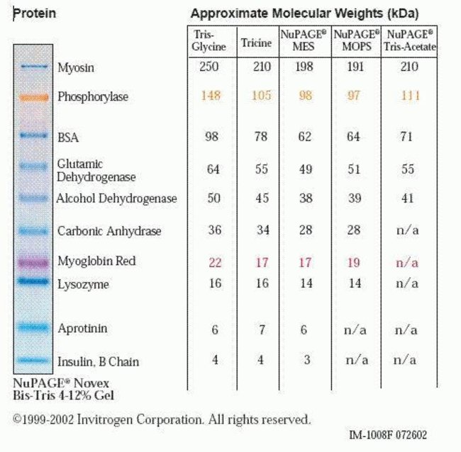
Find additional tips, troubleshooting help, and resources within our Protein Standards and Ladders Support Center.
I used one of your pre-stained protein standards for a western transfer and I noticed that the intensity of the band faded from the membrane during the transfer process. Why is this?
The fading is most likely due to detergent in the western blocking/washing solutions that can remove some of the proteins from the membrane. The dye itself will not wash off of the proteins because it is covalently bound. We have found that smaller pore size membranes retain the proteins better during blocking and wash procedures, and hence recommend use of 0.2 µm instead of 0.45 µm membranes for best resolution and protein retention. After transfer, it is a good idea to circle the pre-stained bands with a pencil on the membrane, so band positions can be identified after blocking and processing.
Find additional tips, troubleshooting help, and resources within our Protein Assays and Analysis Support Center.
I used one of your protein standards for a western transfer and noticed that some of the lower-molecular weight protein bands passed through the membrane. How can I resolve this issue?
- Decrease voltage, current or length of transfer time
- Make sure that the methanol concentration in the transfer buffer is proper; use a methanol concentration of 10-20% methanol removes the SDS from SDS-protein complexes and improves the binding of protein to the membrane.
- Make sure that the SDS concentration (if added) in the transfer buffer is proper, don't use more than 0.02-0.04% SDS. Using too much SDS can prevent binding of proteins to the membrane.
- Check the pore size of the membrane and the size of the target protein. Proteins smaller than 10 kDa will easily pass through a 0.45 µm pore size membrane. If proteins smaller than 10 kDa are of interest, it would be better to use a 0.2 µm pore size membrane.
Find additional tips, troubleshooting help, and resources within our Protein Assays and Analysis Support Center.
I used one of your protein standards for a western transfer and noticed that some of the higher-molecular weight bands transferred very poorly to the membrane. Can you offer some tips?
- Increase voltage, current or length of time for transfer
- SDS in the gel and in the SDS-protein complexes promotes elution of the protein from the gels but inhibits binding of the protein to membranes. This inhibition is higher for nitrocellulose than for PVDF. For proteins that are difficult to elute from the gel such as large molecular weight proteins, a small amount of SDS may be added to the transfer buffer to improve transfer. We recommend pre-equilibrating the gel in 2X Transfer buffer (without methanol) containing 0.02-0.04% SDS for 10 minutes before assembling the sandwich and then transferring using 1X transfer buffer containing 10% methanol and 0.01%SDS.
- Methanol removes the SDS from SDS-protein complexes and improves the binding of protein to the membrane, but has some negative effects on the gel itself, leading to a decrease in transfer efficiency. It may cause a reduction in pore size, precipitation of some proteins, and some basic proteins to become positively charged or neutral. Make sure that the methanol concentration in the transfer buffer is not more than 10-20% and that high-quality, analytical grade methanol is used.
Find additional tips, troubleshooting help, and resources within our Protein Assays and Analysis Support Center.
I used one of your pre-stained standards on a Tris-Glycine gel and noticed that the molecular weights of the proteins were different than on a NuPAGE Bis-Tris gel. What is the reason for this?
Pre-stained standards have a dye that is covalently bound to each protein that will result in the standard migrating differently in different buffer systems (i.e., different gels). As a result, using a pre-stained standard for molecular weight estimation will only give the apparent molecular weight of the protein. Pre-stained standards may be used for molecular weight approximation, confirming gel migration and estimating blotting efficiency but for accurate molecular weight estimation, an unstained standard should be used.
Find additional tips, troubleshooting help, and resources within our Protein Assays and Analysis Support Center.
I used one of your protein standards and am seeing some extra bands in the lane. Can you offer some suggestions?
- While loading, take care to make sure that there is no cross-contamination from adjacent sample lanes.
- Make sure that the correct amount of standard is loaded per lane. Loading too much protein can result in extra bands and this is a problem especially with silver-stained gels.
- Improper storage of the standard or repeated freeze/thawing can result in protein degradation.
Find additional tips, troubleshooting help, and resources within our Protein Assays and Analysis Support Center.
I used one of your protein standards and the bands look non-distinct and smeary. What should I do?
Here are some suggestions:
- Make sure that the correct amount of standard is loaded per lane. Loading too much protein can cause smearing and this is a problem especially with silver stained gels.
- Bands will not be as well resolved in low percentage gels. Try using a higher percentage gel.
- If the bands look smeary and non-distinct after a western transfer/detection, this may be due to the antibody being too concentrated. Follow the manufacturer's recommended dilution or determine the optimal antibody concentration by dot-blotting.
Find additional tips, troubleshooting help, and resources within our Protein Assays and Analysis Support Center.
A couple of bands in my protein standard are missing on the gel. Can you help me troubleshoot?
Here are some suggestions:
- Check the gel type/percentage of the gel that was used. Depending on the gel type and/or percentage, all the bands may not be seen. For example, the smallest bands of the protein standard may not resolve on a very low percentage gel whereas the higher molecular weight bands may not resolve on a high percentage gel.
- Check the expiration date on the protein standard. Expired lots may result in faded or missing bands due to protein degradation.
- Check the storage conditions for the protein standard. Improper storage conditions will compromise the stability of the proteins in the standard.
- Make sure that the protein standard was not heated/boiled prior to loading on the gel. Our protein standards are ready to load and we do not recommend heating/boiling them as this may cause degradation of proteins in the standard.
Find additional tips, troubleshooting help, and resources within our Protein Assays and Analysis Support Center.
Can I combine MagicMark XP Western Protein standard with one of your prestained standards?
You can combine 5 µL MagicMark XP Standard with 5 µL Invitrogen Sharp Prestained Standard or 10 µL MagicMark XP Standard with 5 µL SeeBlue Prestained Standard in the same run using the same lane. The colored bands from the prestained standard can be used to confirm gel run and transfer. Following immunodetection, sharp MagicMark XP bands will develop directly on the western blot.
Find additional tips, troubleshooting help, and resources within our Protein Assays and Analysis Support Center.
What are the storage conditions and shelf life for the SeeBlue Prestained Standard and SeeBlue Plus 2 Prestained Standard?
We recommend storing the SeeBlue Prestained Standard and SeeBlue Plus 2 Prestained Standard at 4 degrees C. They are stable for 4 months when properly stored.
Find additional tips, troubleshooting help, and resources within our Protein Assays and Analysis Support Center.
What is the nature of the dyes that are bound to the proteins in the SeeBlue Prestained Standard and SeeBlue Plus 2 Prestained Standard?
The dyes used are proprietary. All the blue bands in the SeeBlue Prestained Standard (9 blue bands) and SeeBlue Plus 2 Prestained Standard (8 blue bands) have the same dye coupled to them. In addition, the SeeBlue Plus 2 Prestained Standard has two colored bands.
Find additional tips, troubleshooting help, and resources within our Protein Assays and Analysis Support Center.
What are the origins of the proteins in the SeeBlue Prestained Standard and SeeBlue Plus 2 Prestained Standard?
The origins of the proteins in the SeeBlue Prestained Standard and SeeBlue Plus 2 Prestained Standard are as foll:
- Myosin: Rabbit muscle
- BSA: Bovine serum
- Glutamic dehydrogenase: Bovine liver
- Alcohol dehydrogenase: Baker's yeast
- Carbonic anhydrase: Bovine
- Myoglobin: Horse heart
- Lysozyme: Egg white
- Aprotinin: Bovine lung
- Insulin B chain: Bovine pancreas
Find additional tips, troubleshooting help, and resources within our Protein Assays and Analysis Support Center.
I am not sure whether I have the SeeBlue Prestained Standard or SeeBlue Plus 2 Prestained Standard. Is there a way of telling the difference from the cap color?
The SeeBlue Prestained Standard has a blue cap whereas the SeeBlue Plus 2 Prestained Standard has a reddish orange cap.
Find additional tips, troubleshooting help, and resources within our Protein Assays and Analysis Support Center.
Do protein standards run differently on a Zymogram gel compared to a regular Tris-Glycine gel?
Zymogram gels are essentially Tris-Glycine gels containing the substrate. Protein standards run based solely on the percentage of acrylamide and hence should run the same in both kinds of gels. It is quite possible though that if the standard is prestained, the proteins will appear a different color because of the staining (or pre-staining) of the Zymogram gels.
Find additional tips, troubleshooting help, and resources within our Protein Assays and Analysis Support Center.
Can I use any of your protein standards for estimation of protein quantity (protein quantitation)?
Our protein standards are not designed for protein quantitation.
Find additional tips, troubleshooting help, and resources within our Protein Assays and Analysis Support Center.
Can I use your prestained standards for native gel electrophoresis?
We do not recommend using our prestained standards for native gel electrophoresis since they are already denatured (in SDS sample buffer) and pre-reduced (by a proprietary method), and will not resolve well in under native conditions.
Find additional tips, troubleshooting help, and resources within our Protein Assays and Analysis Support Center.
Can I use a prestained protein ladder to estimate the molecular weight of my protein?
We recommend using unstained protein ladders for molecular weight estimation applications as prestained ladders have a dye that is covalently bound to each protein that will result in the ladder migrating differently in different buffer systems (i.e., different gels). As a result, using a prestained ladder for molecular weight estimation will only give the apparent molecular weight of the protein. Prestained ladders may be used for molecular weight approximation, confirming gel migration and estimating blotting efficiency but for accurate molecular weight estimation, an unstained ladder should be used.
Find additional tips, troubleshooting help, and resources within our Protein Assays and Analysis Support Center.
What is the recommended gel loading volume for your protein standards?
Please find this information in the respective manuals for the individual protein standards.
Find additional tips, troubleshooting help, and resources within our Protein Standards and Ladders Support Center.
Do I need to boil your protein standards before loading on the gel?
Our protein standards are ready to load. We do not recommend heating them as this may cause protein degradation.
Find additional tips, troubleshooting help, and resources within our Protein Assays and Analysis Support Center.
Do I have to add reducing agent to your protein standards?
Except for our NativeMark Unstained Protein Standard (designed for native electrophoresis), all of the other unstained and prestained standards we offer (Invitrogen Sharp, SeeBlue, SeeBlue Plus2, BenchMark, HiMark) have been pre-reduced (by a proprietary method). Hence, you do not need to add reducing agent.
Find additional tips, troubleshooting help, and resources within our Protein Assays and Analysis Support Center.
Which pre-stained protein standards are compatible with Bolt Bis-Tris Plus gels?
SeeBlue prestained standard, SeeBlue Plus 2 prestained standard and Invitrogen Sharp prestained standard are compatible with Bolt Bis-Tris Plus gels. Their migration is the same as that in NuPAGE gels with the corresponding buffer.
Find additional tips, troubleshooting help, and resources within our Protein Electrophoresis and Western Blotting Support Center.