Search


Citations & References (4)
Invitrogen™
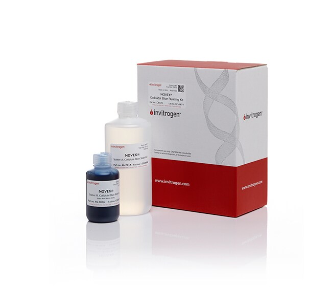
Colloidal Blue Staining Kit
The Colloidal Blue Staining Kit allows detection of nanogram levels of proteins in 1D or 2D PAGE gels with minimalRead more
| Catalog Number | Quantity |
|---|---|
| LC6025 | 1 kit |
Catalog number LC6025
Price (CLP)
375.569
Each
Quantity:
1 kit
Price (CLP)
375.569
Each
The Colloidal Blue Staining Kit allows detection of nanogram levels of proteins in 1D or 2D PAGE gels with minimal effort and requires only water to destain. The Colloidal Blue stain uses colloidal chemistry that reduces free dye in solution and improves the protein-to-dye binding ratio. Samples are intensely stained and visible within three hours. Background staining is virtually eliminated by destaining overnight with water. The kit requires one easy solution preparation. Methanol is required in the staining step, but is not included in the kit.
Compare all Coomassie stains ›
Compare all Coomassie stains ›
For Research Use Only. Not for use in diagnostic procedures.
Specifications
Detection LocationIn-Gel Detection
Detection MethodColorimetric
Quantity1 kit
Shelf Life6 Months
Shipping ConditionRoom Temperature
Target MoleculeProtein
Label or DyeCoomassie
Product TypeProtein Gel Stain Kit
Unit SizeEach
Contents & Storage
The Colloidal Blue Staining Kit is supplied with sufficient stainer A and stainer B to stain 25 mini-gels. Store the kit at room temperature. The kit is guaranteed for 6 months when properly stored unless otherwise stated in the product documentation.
Frequently asked questions (FAQs)
Can I use the Colloidal Blue Staining Kit (Cat. No. LC6025) for membrane staining to detect proteins that are transferred to a membrane?
Why is Coomassie G-250 used as the tracking dye in NuPAGE LDS Sample Buffer instead of bromophenol blue?
Is the SimplyBlue SafeStain the appropriate stain for quantitation by densitometry?
The sensitivity of my SimplyBlue SafeStain seems to be decreasing over time. Why is this?
How do I destain proteins on a PVDF membrane that were stained with SimplyBlue SafeStain?
Citations & References (4)
Citations & References
Abstract
SALSA, a variant of yeast SAGA, contains truncated Spt7, which correlates with activated transcription.
Journal:Proc Natl Acad Sci U S A
PubMed ID:12186975
'Spt-Ada-Gcn5 acetyltransferase (SAGA) is a previously described histone acetyltransferase/transcriptional coactivator complex in yeast. At promoters of certain genes (HIS3 and TRP3), SAGA has an inhibitory function involving a nonproductive TATA-binding protein interaction mediated by the Spt3 and Spt8 subunits. Related to this, Spt8-less SAGA is a major form of the
Chromatin deacetylation by an ATP-dependent nucleosome remodelling complex.
Journal:Nature
PubMed ID:9804427
'The dynamic assembly and remodelling of eukaryotic chromosomes facilitate fundamental cellular processes such as DNA replication and gene transcription. The repeating unit of eukaryotic chromosomes is the nucleosome core, consisting of DNA wound about a defined octamer of histone proteins. Two enzymatic processes that regulate transcription by targeting elements of
A family of chromatin remodeling factors related to Williams syndrome transcription factor
Journal:Proc Natl Acad Sci U S A
PubMed ID:10655480
Chromatin remodeling complexes have been implicated in the disruption or reformation of nucleosomal arrays resulting in modulation of transcription, DNA replication, and DNA repair. Here we report the isolation of WCRF, a new chromatin-remodeling complex from HeLa cells. WCRF is composed of two subunits, WCRF135, the human homolog of Drosophila
A method to identify serine kinase substrates. Akt phosphorylates a novel adipocyte protein with a Rab GTPase-activating protein (GAP) domain.
Journal:J Biol Chem
PubMed ID:11994271
This study describes a method for the identification of the substrates of specific serine kinases. An antibody specific for the phosphomotif generated by the kinase is used to isolate phosphorylated substrates by immunoprecipitation, and the isolated proteins are identified by tandem mass spectrometry of peptides. This method was applied to