Search
Zeta
CD22 Monoclonal Antibody (ZM183), MonoMab™
{{$productOrderCtrl.translations['antibody.pdp.commerceCard.promotion.promotions']}}
{{$productOrderCtrl.translations['antibody.pdp.commerceCard.promotion.viewpromo']}}
{{$productOrderCtrl.translations['antibody.pdp.commerceCard.promotion.promocode']}}: {{promo.promoCode}} {{promo.promoTitle}} {{promo.promoDescription}}. {{$productOrderCtrl.translations['antibody.pdp.commerceCard.promotion.learnmore']}}
Additional Information:
{{banner.description}}
Product Details
Z2496MP
Applications
Tested Dilution
Publications
Product Specifications
Species Reactivity
Human
Host/Isotype
Mouse
/ IgG1, kappa
Class
Monoclonal
Type
Antibody
Clone
ZM183
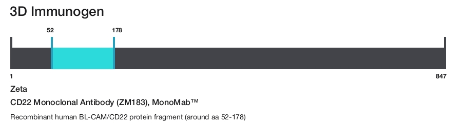
Immunogen
Recombinant human BL-CAM/CD22 protein fragment (around aa 52-178)
Conjugate
Form
Liquid
Purification
Protein A
Storage buffer
tris with NP-40, BSA
Contains
<0.1% sodium azide
Storage conditions
4°C
Shipping conditions
Ambient (domestic); Wet ice (international)
Product Specific Information
This product is diluted and in a ready-to-use formulation.
A recommended positive control tissue for this product is Lymph Node, however positive controls are not limited to this tissue type.
The primary antibody is intended for laboratory professional use in the detection of the corresponding protein in formalin-fixed, paraffin-embedded tissue stained in manual qualitative immunohistochemistry (IHC) testing. This antibody is intended to be used after the primary diagnosis of tumor has been made by conventional histopathology using non-immunological histochemical stains.
Recognizes a protein of 130-140 kDa, identified as CD22 (also known as BL-CAM). CD22 expression is restricted to normal and neoplastic B cells and is absent from other haemopoietic cell types. In B-cell ontogeny, CD22 is first expressed in the cytoplasm of pro-B and pre-B cells, and on the surface as B cells mature to become IgD+. It is not expressed by plasma cells, CD22 is found highly expressed in follicular mantle and marginal zone B-cells, and while germinal center B-cells are relatively weak. CD22 is a member of the immunoglobulin superfamily and serves as an adhesion receptor for sialic acid-bearing ligands expressed on erythrocytes and all leukocyte classes. It also associates with tyrosine kinases and play a role in signal transduction and B-cell activation.
Antibody is used with formalin-fixed and paraffin-embedded sections. Pretreatment of deparaffinized tissue with heat-induced epitope retrieval or enzymatic retrieval is recommended. In general, immunohistochemical (IHC) staining techniques allow for the visualization of antigens via the sequential application of a specific antibody to the antigen (primary antibody), a secondary antibody to the primary antibody (link antibody), an enzyme complex and a chromogenic substrate with interposed washing steps. The enzymatic activation of the chromogen results in a visible reaction product at the antigen site. Results are interpreted using a light microscope and aid in the differential diagnosis of pathophysiological processes, which may or may not be associated with a particular antigen.
A positive tissue control must be run with every staining procedure performed. This tissue may contain both positive and negative staining cells or tissue components and serve as both the positive and negative control tissue. External Positive control materials should be fresh autopsy/biopsy/surgical specimens fixed, processed and embedded as soon as possible in the same manner as the patient sample (s). Positive tissue controls are indicative of correctly prepared tissues and proper staining methods. The tissues used for the external positive control materials should be selected from the patient specimens with well-characterized low levels of the positive target activity that gives weak positive staining. The low level of positivity for external positive controls is designed to ensure detection of subtle changes in the primary antibody sensitivity from instability or problems with the staining methodology. A tissue with weak positive staining is more suitable for optimal quality control and for detecting minor levels of reagent degradation.
Internal or external negative control tissue may be used depending on the guidelines and policies that govern the organization to which the end user belongs to. The variety of cell types present in many tissue sections offers internal negative control sites, but this should be verified by the user. The components that do not stain should demonstrate the absence of specific staining, and provide an indication of non-specific background staining. If specific staining occurs in the negative tissue control sites, results with the patient specimens must be considered invalid.
Target Information
CD22, also known as BL-CAM, is a type I transmembrane glycoprotein composed of two polypeptide chains, CD22alpha and CD22beta, with molecular weights of 130 and 140 kDa, respectively. These chains are produced by alternative splicing of the CD22 gene. CD22 is prominently expressed on mature B cells and B cell lymphomas, including hairy cell leukemia, diffuse large B-cell lymphoma, and nodular lymphocyte predominance Hodgkin's lymphoma, but is negative in classical Hodgkin's lymphoma. The extracellular portion of CD22 contains seven Ig-like domains that preferentially bind alpha2,6-linked sialic acid moieties found on epithelial, endothelial, B, and T cells. This binding can be masked by cis interactions with sialic acids on the same cell surface. CD22 expression is limited to late stages of B-cell differentiation, making it useful for phenotyping mature leukemias. Intracellularly, CD22 features six tyrosine residues within immunotyrosine-based inhibitory motifs (ITIM) and activation-like motifs. These residues are phosphorylated upon B-cell receptor engagement, allowing CD22 to regulate B-cell receptor signaling. CD22 participates in positive regulation through interactions with Src family tyrosine kinases and acts as an inhibitory receptor by recruiting cytoplasmic phosphatases via SH2 domains, which block signal transduction through dephosphorylation of signaling molecules. CD22's role in both positive and negative regulation of B-cell signaling, along with its specific expression pattern, makes it a valuable marker for antibody customers interested in B-cell-related research and diagnostics.
For Research Use Only. Not for use in diagnostic procedures. Not for resale without express authorization.
References (0)
Have you cited this product in a publication?
Let us know so we can reference it here.
Bioinformatics
Protein Aliases: B-cell receptor CD22; B-lymphocyte cell adhesion molecule; B-lymphocyte cell adhesion molecule (BL-CAM); BL-CAM; CD22; CD22 antigen; FLJ22814; Lectin 2; Leu-14; MGC130020; Sialic acid-binding Ig-like lectin 2; Sialic acid-binding Ig-like lectin 2 (Siglec-2); Siglec-2; T-cell surface antigen Leu-14; unnamed protein product
Gene Aliases: CD22; SIGLEC-2; SIGLEC2
UniProt ID: (Human) P20273
Entrez Gene ID: (Human) 933
negative regulation of immunoglobulin production
negative regulation of immune system process
cell adhesion
negative regulation of signal transduction
regulation of endocytosis
regulation of B cell proliferation
B cell activation
regulation of immune response
negative regulation of calcium-mediated signaling
negative regulation of B cell receptor signaling pathway
Disclaimer

Clicking the images or links will redirect you to a website hosted by BenchSci that provides third-party scientific content. Neither the content nor the BenchSci technology and processes for selection have been evaluated by us; we are providing them as-is and without warranty of any kind, including for use or application of the Thermo Fisher Scientific products presented.

Performance Guarantee
If an Invitrogen™ antibody doesn't perform as described on our website or datasheet,we'll replace the product at no cost to you, or provide you with a credit for a future purchase.*
Learn more
We're here to help
Get expert recommendations for common problems or connect directly with an on staff expert for technical assistance related to applications, equipment and general product use.
Contact tech support