Search
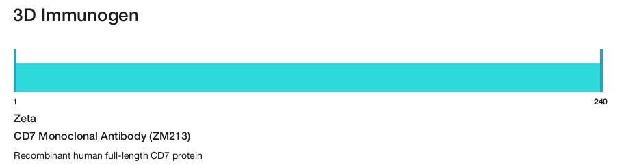
Zeta
CD7 Monoclonal Antibody (ZM213)
{{$productOrderCtrl.translations['antibody.pdp.commerceCard.promotion.promotions']}}
{{$productOrderCtrl.translations['antibody.pdp.commerceCard.promotion.viewpromo']}}
{{$productOrderCtrl.translations['antibody.pdp.commerceCard.promotion.promocode']}}: {{promo.promoCode}} {{promo.promoTitle}} {{promo.promoDescription}}. {{$productOrderCtrl.translations['antibody.pdp.commerceCard.promotion.learnmore']}}
Additional Information:
{{banner.description}}
Product Details
Z2548MP
Applications
Tested Dilution
Publications
Product Specifications
Species Reactivity
Human
Host/Isotype
Mouse
/ IgG1, kappa
Class
Monoclonal
Type
Antibody
Clone
ZM213
Immunogen
Recombinant human full-length CD7 protein
Conjugate
Form
Liquid
Purification
Protein A
Storage buffer
tris with NP-40, BSA
Contains
<0.1% sodium azide
Storage conditions
4°C
Shipping conditions
Ambient (domestic); Wet ice (international)
Product Specific Information
This product is diluted and in a ready-to-use formulation.
A recommended positive control tissue for this product is Lymph Node, however positive controls are not limited to this tissue type.
The primary antibody is intended for laboratory professional use in the detection of the corresponding protein in formalin-fixed, paraffin-embedded tissue stained in manual qualitative immunohistochemistry (IHC) testing. This antibody is intended to be used after the primary diagnosis of tumor has been made by conventional histopathology using non-immunological histochemical stains.
CD7 is a single-pass type 1 transmembrane protein that is a member of the immunoglobulin superfamily. It plays an essential role in T-cell interactions and also in T-cell/B-cell interactions during early lymphoid development. CD7 is expressed on thymocytes, T-and natural killer cells, and progenitors of lymphoid and myeloid cells. It is also expressed on T-cell Acute Lymphoblastic Leukemia/Lymphoma, Acute Myelogenous Leukemia and Chronic Myelogenous Leukemia. CD7 antibody is the most sensitive and specific T-cell deletion marker. Loss of CD7 expression by neoplastic lymphocytes is considered a distinguishing characteristic of mycosis fungoides (MF) and cutaneous T-cell lymphoma.
Antibody is used with formalin-fixed and paraffin-embedded sections. Pretreatment of deparaffinized tissue with heat-induced epitope retrieval or enzymatic retrieval is recommended. In general, immunohistochemical (IHC) staining techniques allow for the visualization of antigens via the sequential application of a specific antibody to the antigen (primary antibody), a secondary antibody to the primary antibody (link antibody), an enzyme complex and a chromogenic substrate with interposed washing steps. The enzymatic activation of the chromogen results in a visible reaction product at the antigen site. Results are interpreted using a light microscope and aid in the differential diagnosis of pathophysiological processes, which may or may not be associated with a particular antigen.
A positive tissue control must be run with every staining procedure performed. This tissue may contain both positive and negative staining cells or tissue components and serve as both the positive and negative control tissue. External Positive control materials should be fresh autopsy/biopsy/surgical specimens fixed, processed and embedded as soon as possible in the same manner as the patient sample (s). Positive tissue controls are indicative of correctly prepared tissues and proper staining methods. The tissues used for the external positive control materials should be selected from the patient specimens with well-characterized low levels of the positive target activity that gives weak positive staining. The low level of positivity for external positive controls is designed to ensure detection of subtle changes in the primary antibody sensitivity from instability or problems with the staining methodology. A tissue with weak positive staining is more suitable for optimal quality control and for detecting minor levels of reagent degradation.
Internal or external negative control tissue may be used depending on the guidelines and policies that govern the organization to which the end user belongs to. The variety of cell types present in many tissue sections offers internal negative control sites, but this should be verified by the user. The components that do not stain should demonstrate the absence of specific staining, and provide an indication of non-specific background staining. If specific staining occurs in the negative tissue control sites, results with the patient specimens must be considered invalid.
Target Information
CD7, also known as gp40 or Leu9, is a 40 kDa receptor and member of the immunoglobulin gene superfamily. It features an N-terminal region (amino acids 1-107) that is highly homologous to Ig kappa light chains, while its carboxyl-terminal region is proline-rich, forming a stalk from which the Ig domain projects. CD7 is prominently expressed on the majority of immature and mature T lymphocytes, as well as T cell leukemias. It is also found on natural killer cells, a small subpopulation of normal B cells, and malignant B cells. CD7 plays a crucial role in modulating immune cell activity. Cross-linking of surface CD7 enhances T cell and NK cell functions, as evidenced by increased calcium flux, expression of adhesion molecules, cytokine secretion, and proliferation. CD7 directly associates with phosphoinositol 3-kinase, and its ligation induces the production of D-3 phosphoinositides and tyrosine phosphorylation. The expression of CD7 is an important marker in leukemia diagnostics, highlighting its significance in both normal immune function and disease states.
For Research Use Only. Not for use in diagnostic procedures. Not for resale without express authorization.
References (0)
Have you cited this product in a publication?
Let us know so we can reference it here.
Bioinformatics
Protein Aliases: CD7; CD7 antigen (p41); GP40; p41 protein; T-cell antigen CD7; T-cell leukemia antigen; T-cell surface antigen Leu-9; TP41; unnamed protein product
Gene Aliases: CD7; GP40; LEU-9; Tp40; TP41
UniProt ID: (Human) P09564
Entrez Gene ID: (Human) 924
Disclaimer

Clicking the images or links will redirect you to a website hosted by BenchSci that provides third-party scientific content. Neither the content nor the BenchSci technology and processes for selection have been evaluated by us; we are providing them as-is and without warranty of any kind, including for use or application of the Thermo Fisher Scientific products presented.

Performance Guarantee
If an Invitrogen™ antibody doesn't perform as described on our website or datasheet,we'll replace the product at no cost to you, or provide you with a credit for a future purchase.*
Learn more
We're here to help
Get expert recommendations for common problems or connect directly with an on staff expert for technical assistance related to applications, equipment and general product use.
Contact tech support