Search


Citas y referencias (16)
Invitrogen™
pHrodo™ BioParticles™ Conjugates for Phagocytosis and Phagocytosis Kit, for Flow Cytometry
Obtenga resultados más rápidos y exactos en ensayos de fagocitosis con los conjugados y el kit pHrodo BioParticles. Los conjugados pHrodo BioParticles muestran una fluorescencia brillante en los endosomas y lisosomas, no requieren pasos de lavado ni colorantes supresores, se pueden multiplexar y se utilizan en citometría de flujo, HCA y HTS.
Have Questions?
Cambiar vista

| Número de catálogo | Tipo de producto | Especie | Color |
|---|---|---|---|
| P35365 | Conjugado | S. cerevisiae | |
| P35360 | Conjugate | E. coli | |
| P35361 | Conjugado | E. coli | |
| A10025 | Kit de fagocitosis | E. coli | |
| A10010 | Conjugado | S. aureus | |
| P35367 | Conjugado | S. aureus |
Número de catálogo P35365
Precio (USD)
926,64
Each
Tipo de producto:
Conjugado
Especie:
S. cerevisiae
Precio (USD)
926,64
Each
Consiga una tinción más rápida y resultados más exactos sin necesidad de pasos de lavado o colorantes supresores con conjugados pHrodo BioParticles sensibles al pH y kit de fagocitosis para citometría de flujo. Los conjugados pHrodo BioParticles para fagocitosis no son fluorescentes fuera de la celda con pH neutro, pero muestran fluorescencia brillante en ambientes con pH ácido, como los de los endosomas y lisosomas. Se pueden utilizar en aplicaciones de adquisición de imágenes, HCA y HTA. El kit de fagocitosis pHrodo BioParticles ofrece una evaluación rápida de la actividad fagocítica en sangre completa mediante citometría de flujo.
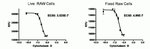
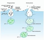
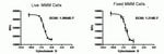
The fluorescence of pHrodo dye dramatically increases as pH decreases from neutral to acidic, making it an ideal tool to study phagocytosis and its regulation by drugs and/or environmental factors. Outside the cell, the dye is non-fluorescent, which eliminates the need for wash steps and quencher dyes. Once internalized, the dye fluoresces brightly within the acidic environments of endosomes and lysosomes, enabling rapid assay development and certainty of results when investigating phagocytic pathways and their regulation by drugs and/or environmental factors. As such, pHrodo dye conjugates can be used in plate readers, fluorescence microscopy imaging, and flow cytometry applications.
The pHrodo Red S. aureus BioParticles Conjugate for Phagocytosis (Cat. No. A10010) offers fast and accurate assay results. pHrodo Red dye conjugates are non-fluorescent outside the cell but fluoresce bright red in phagosomes. The pHrodo Red E. coli BioParticles Conjugate for Phagocytosis (Cat. No. P35361) detects phagocytosis and endocytosis, offers reduced signal variability and improved timing in sensitive experiments, and can be used in multiplex applications with a wide variety of blue, green, and far-red dyes and reporters such as GFP, Fuo-4, calcein, NucBlue, CellEvent Caspase 3/7 green, Mitosox Green, and Mitotracker Deep Red, among many others. The optimal absorption and fluorescence emission maxima of the pHrodo Red BioParticles Conjugate is approximately 560/585 nm, respectively. However, the fluorophore is readily excited with the 488-nm argon-ion laser installed on most flow cytometers.
The pHrodo Red E. coli BioParticles Phagocytosis Kit (Cat. No. A10025) for flow cytometry is designed for rapid and convenient measurement of phagocytic activity in whole blood samples by flow cytometry. The kit includes all the reagents required for assessing particle ingestion and red blood cell lysis.
pHrodo Deep Red dye is a low-background pH sensor dye that shows no signal in neutral conditions and only fluoresces in acidic environments. pHrodo Deep Red dye enables better discrimination of internalized cargo from outside the cell because it has an approximate pKa of 5 and does not fluoresce until it enters the late endosome and lysosome. pHrodo Deep Red dye can be detected using a Cy5 fluorescent filter set and has been validated for use in a variety of applications, including flow cytometry, fluorescent microscopy, high content analysis (HCA), and high throughput screening (HTS). It can also be multiplexed with a wide variety of blue, green, and red dyes and reporters such as GFP and RFPs, Mitosox Green or Red, CellEvent Caspase 3/7 Green or Red, NucBlue, or TMR, among many others.
pHrodo Deep Red E. coli BioParticles Conjugate for Phagocytosis (Cat. No. P35360) is a no-wash, low background, fluorogenic reagent developed for the study of phagocytosis in a live cell system. This conjugate is non-fluorescent outside the cell but fluoresces dark red in phagosomes. The low pKa of the pHrodo Deep Red E. coli BioParticles Conjugate eliminates non-specific signal from non-internalized cargo and only turns on in late endosomes and lysosomes.
The pHrodo Green dye conjugates, like pHrodo Red and pHrodo Deep Red dye conjugates, offer faster and more accurate results than any other phagocytosis assay. pHrodo Green conjugates are non-fluorescent outside the cell at neutral pH but fluoresce bright green at acidic pH, such as that of phagosomes. Use the ready-made pHrodo Green S. aureus BioParticles Conjugate (Cat. No. P35367) and pHrodo Green Zymosan BioParticles Conjugate (Cat. No. P35365) for Phagocytosis in imaging, HCA, HTS, and flow applications.
These pHrodo conjugate products include enough reagent to perform 100 tests when using 100 μL in each well of a 96-well plate, and they can be multiplexed with a wide variety of blue, red, and far-red dye reporters such as Mitosox Red, CellEvent Caspase 3/7 Red, NucBlue, RFPs, and Mitotracker Deep Red, among many others.
pHrodo Red Zymosan BioParticles Conjugate (Cat. No. P35364) is also available, and pHrodo Green dye is available as a conjugate of E. coli BioParticles (Cat. No. P35366).
For Research Use Only. Not for human or animal therapeutic or diagnostic use.
Especificaciones
DescripciónConjugado para fagocitosis pHrodo™ Green Zymosan BioParticles™
Método de detecciónFluorescente
Tipo de coloranteColoraciones sensibles al pH
Excitación/emisión509/533
Cantidad5 x 1 mg
Condiciones de envíoTemperatura ambiente
EspecieS. cerevisiae
ColorVerde
Para utilizar con (aplicación)Análisis celular
Para utilizar con (equipo)Citómetro de focalización acústica Attune™, Microscopio confocal, Sistema de adquisición de imágenes de células Floid™, Microscopio de fluorescencia, Instrumentos de alto contenido, Citómetro de flujo
Línea de productospHrodo
Tipo de productoConjugado
Unit SizeEach
Contenido y almacenamiento
Almacenar a – 20 °C, desecar y proteger de la luz.
Preguntas frecuentes
Can I store reconstituted pHrodo BioParticles Conjugates for Phagocytosis and Phagocytosis Kit, for Flow Cytometry?
Are the Invitrogen BioParticles products sterile?
What is the type of bond that attaches the dyes to the BioParticles probes?
What cellular processes can be analyzed with a flow cytometer?
What can the BioParticles product line be used for?
Citations & References (16)
Citations & References
Abstract
A Review of Reagents for Fluorescence Microscopy of Cellular Compartments and Structures, Part I: BacMam Labeling and Reagents for Vesicular Structures.
Journal:
PubMed ID:23835803
'Fluorescent labeling of vesicular structures in cultured cells, particularly for live cells, can be challenging for a number of reasons. The first challenge is to identify a reagent that will be specific enough where some structures have a number of potential reagents and others very few options. The emergence of
A modern descendant of early green algal phagotrophs.
Journal:
PubMed ID:23707430
Green algae, land plants, and other photosynthetic eukaryotes possess plastids, such as chloroplasts, which have evolved from cyanobacterial ancestors via endosymbiosis [1]. An early evolutionary merger between heterotrophic eukaryotes and cyanobacteria called primary endosymbiosis gave rise to the first photosynthetic eukaryotes. A series of plastid acquisitions involving engulfment of eukaryotic phototrophs,
Glutathione reductase is essential for host defense against bacterial infection.
Journal:Free Radic Biol Med
PubMed ID:23623936
Glutathione reductase (Gsr) catalyzes the reduction of glutathione disulfide to glutathione, a major cellular antioxidant. We have recently shown that Gsr is essential for host defense against the gram-negative bacteria Escherichia coli in a mouse model of sepsis. Although we have demonstrated that Gsr is required for sustaining the oxidative
NFAT signaling in human mesenchymal stromal cells affects extracellular matrix remodeling and antifungal immune responses.
Journal:iScience
PubMed ID:34195564
Deficiency of Microglial Autophagy Increases the Density of Oligodendrocytes and Susceptibility to Severe Forms of Seizures.
Journal:Eneuro
PubMed ID:33472865