Search

Invitrogen™
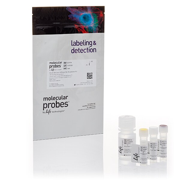
Pacific Blue™ Annexin V/SYTOX™ AADvanced™ Apoptosis Kit, for flow cytometry
Ce produit détecte l’externalisation de la phosphatidylsérine dans les cellules apoptotiques à l’aide d’annexine V recombinante conjuguée au colorant violetAfficher plus
| Référence | Quantité |
|---|---|
| A35136 | 1 kit |
Référence A35136
Prix (EUR)
524,00
Each
Quantité:
1 kit
Prix (EUR)
524,00
Each
Ce produit détecte l’externalisation de la phosphatidylsérine dans les cellules apoptotiques à l’aide d’annexine V recombinante conjuguée au colorant violet fluorescent Pacific Blue™ et dans les cellules mortes à l’aide de colorant SYTOX™ AADvanced™. Après avoir coloré une population cellulaire avec du Pacific Blue™, de l’annexine V et du SYTOX™ AADvanced™, les cellules apoptotiques présentent une fluorescence violette, les cellules mortes une fluorescence rouge et les cellules vivantes une fluorescence faible ou nulle. Ces populations sont facilement distinguées par un cytomètre en flux avec les lignes de 405 nm et 488 nm pour l’excitation. Il y a très peu de chevauchement spectral entre les deux colorants, ce qui ne nécessite alors qu’une très légère compensation. Chaque kit contient suffisamment de réactifs pour environ 50 tests de cytométrie en flux.
Consultez un guide de sélection pour tous les dosages d’apoptose pour la cytométrie en flux.
Consultez un guide de sélection pour tous les dosages d’apoptose pour la cytométrie en flux.
Usage exclusivement réservé à la recherche. Ne pas utiliser pour des procédures de diagnostic.
Spécifications
Excitation / émissionPacific Blue™ : 415⁄455, SYTOX™ AADvanced™ : 546 / 647
Faisceaux laser du cytomètre en flux405, 488
À utiliser avec (application)Cytométrie en flux
À utiliser avec (équipement)Cytomètre en flux
Nbre de réactions50
Type de produitKit d’apoptose
Quantité1 kit
Conditions d’expéditionGlace humide
ConjuguéColoration pour cellules mortes Pacific Blue™, SYTOX™ AADvanced™
FormatTube
Unit SizeEach
Contenu et stockage
Contient 1 flacon d’annexine V, conjugué Pacific Blue™ (250 µl), 1 flacon de coloration de cellules mortes SYTOX™ AADvanced™ et 1 bouteille de tampon de liaison à l’annexine (solution 5X, 15 ml) et 1 flacon de DMSO (100 µl).
Conserver au réfrigérateur (2–8°C) et à l’abri de la lumière.
Conserver au réfrigérateur (2–8°C) et à l’abri de la lumière.