Search


Citations et références (3)
Invitrogen™
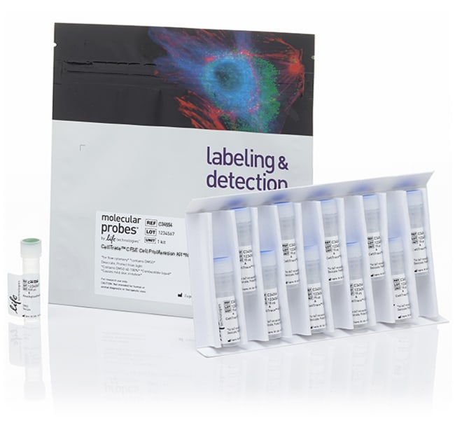
Kit de prolifération cellulaire CellTrace™ CFSE pour cytométrie en flux
Le kit de prolifération cellulaire CFSE CellTrace™ est utilisé pour le marquage in vitro et in vivo des cellules afinAfficher plus
Have Questions?
Modifier l'affichage

| Référence | Suffisant pour |
|---|---|
| C34554 | 180 réactions |
| C34570 | 20 réactions |
Référence C34554
Prix (EUR)
278,65
Offre exceptionnelle en ligne
332,00Économisez 53,35 (16%)
Each
Suffisant pour:
180 réactions
Prix (EUR)
278,65
Offre exceptionnelle en ligne
332,00Économisez 53,35 (16%)
Each
Le kit de prolifération cellulaire CFSE CellTrace™ est utilisé pour le marquage in vitro et in vivo des cellules afin de tracer plusieurs générations à l’aide de la dilution de colorants par cytométrie de flux.
• Performance supérieure—la coloration vive à pic simple permet de visualiser plusieurs générations
• Stabilité du signal à long terme—bien retenu dans les cellules pendant plusieurs jours après la coloration
• Non toxique—la coloration n’a pas d’effet nocif sur la santé des cellules
• Protocole de coloration simple et robuste
Consultez un guide de sélection pour tous™ les kits de prolifération cellulaire CellTrace pour la cytométrie en flux.
Coloration fluorescente supérieure
Une analyse de prolifération réussie par dilution de colorant (voir la figure ci-dessous) nécessite un colorant extrêmement lumineux pour distinguer les cellules marquées par fluorescence de l’auto-fluorescence après plusieurs divisions cellulaires. La coloration fluorescente intense procurée par les colorants CellTrace™ CFSE permet la visualisation de huit générations ou plus de cellules proliférant avant que le signal soit submergé par l’auto-fluorescence intrinsèque des cellules. Une coloration uniforme et homogène entraîne très peu de variation de fluorescence entre les cellules d’une population, de sorte que des générations distinctes peuvent être observées sans aucune exigence de logiciel de modélisation complexe.
Rétention du signal à long terme
Contrairement aux colorations qui marquent la membrane lipidique des cellules, le colorant CellTrace™ CFSE traverse facilement la membrane du plasma et se lie de façon covalente à l’intérieur des cellules où le colorant fluorescent, bien retenu et stable, est conçu pour donner un signal constant, même après plusieurs jours dans un environnement de culture cellulaire.
Non toxique
Le colorant CellTrace™ CFSE se lie de façon covalente à toutes les amines libres à la surface et à l’intérieur des cellules et montre peu de cytotoxicité, avec un effet minimal observé sur la capacité de prolifération ou sur la biologie des cellules. Les chercheurs ont utilisé le marquage CFSE SE pour montrer que les cellules hématopoïétiques transplantables prolifèrent in vitro en réponse à une stimulation par un cocktail de facteur de croissance.
Protocole de coloration simple et robuste
Le kit de prolifération cellulaire CellTrace™ contient des flacons de colorant sec jetables pratiques pour permettre de réaliser des expériences à petite échelle sans préparer des quantités excessives de colorant. Une solution mère est préparée en dissolvant le contenu d’un flacon dans du diméthylsulfoxyde (DMSO) anhydre avant l’utilisation. Pour colorer 1 ml de cellules dans un milieu de culture exempt de protéines, il suffit en général d’utiliser 1 µl de cette solution mère. Les cellules doivent être colorées pendant 20 minutes à température ambiante en agitant doucement. Un bref lavage avec du milieu complet pourra alors absorber tout colorant restant dans la solution.
• Performance supérieure—la coloration vive à pic simple permet de visualiser plusieurs générations
• Stabilité du signal à long terme—bien retenu dans les cellules pendant plusieurs jours après la coloration
• Non toxique—la coloration n’a pas d’effet nocif sur la santé des cellules
• Protocole de coloration simple et robuste
Consultez un guide de sélection pour tous™ les kits de prolifération cellulaire CellTrace pour la cytométrie en flux.
Coloration fluorescente supérieure
Une analyse de prolifération réussie par dilution de colorant (voir la figure ci-dessous) nécessite un colorant extrêmement lumineux pour distinguer les cellules marquées par fluorescence de l’auto-fluorescence après plusieurs divisions cellulaires. La coloration fluorescente intense procurée par les colorants CellTrace™ CFSE permet la visualisation de huit générations ou plus de cellules proliférant avant que le signal soit submergé par l’auto-fluorescence intrinsèque des cellules. Une coloration uniforme et homogène entraîne très peu de variation de fluorescence entre les cellules d’une population, de sorte que des générations distinctes peuvent être observées sans aucune exigence de logiciel de modélisation complexe.
Rétention du signal à long terme
Contrairement aux colorations qui marquent la membrane lipidique des cellules, le colorant CellTrace™ CFSE traverse facilement la membrane du plasma et se lie de façon covalente à l’intérieur des cellules où le colorant fluorescent, bien retenu et stable, est conçu pour donner un signal constant, même après plusieurs jours dans un environnement de culture cellulaire.
Non toxique
Le colorant CellTrace™ CFSE se lie de façon covalente à toutes les amines libres à la surface et à l’intérieur des cellules et montre peu de cytotoxicité, avec un effet minimal observé sur la capacité de prolifération ou sur la biologie des cellules. Les chercheurs ont utilisé le marquage CFSE SE pour montrer que les cellules hématopoïétiques transplantables prolifèrent in vitro en réponse à une stimulation par un cocktail de facteur de croissance.
Protocole de coloration simple et robuste
Le kit de prolifération cellulaire CellTrace™ contient des flacons de colorant sec jetables pratiques pour permettre de réaliser des expériences à petite échelle sans préparer des quantités excessives de colorant. Une solution mère est préparée en dissolvant le contenu d’un flacon dans du diméthylsulfoxyde (DMSO) anhydre avant l’utilisation. Pour colorer 1 ml de cellules dans un milieu de culture exempt de protéines, il suffit en général d’utiliser 1 µl de cette solution mère. Les cellules doivent être colorées pendant 20 minutes à température ambiante en agitant doucement. Un bref lavage avec du milieu complet pourra alors absorber tout colorant restant dans la solution.
Usage exclusivement réservé à la recherche. Ne pas utiliser pour des procédures de diagnostic.
Spécifications
DescriptionKit de prolifération cellulaire CellTrace™, 180 rxns, CFSE, lyophilisé, méthode de détection fluorescente, longueur d’onde d’excitation/émission de 492/517 nm, étiquette / colorant CellTrace™ CFSE, stockage de –5 à –30°C, à utiliser avec le cytomètre de flux
Méthode de détectionFluorescence
Type de colorantAutre(s) étiquette(s) ou colorant(s)
FormeLyophilisé(e)
FormatTube(s)
Quantité1 kit(s)
Type de réactifCFSE & composés associés
Conditions d’expéditionTempérature ambiante
SolubilitéDMSO (diméthylsulfoxyde)
Suffisant pour180 réactions
Gamme de longueur d’onde d’émission492⁄517
À utiliser avec (application)Test de prolifération, Cytométrie en flux
À utiliser avec (équipement)Cytomètre en flux
Gamme de produitsCellTrace
Type de produitKit de prolifération cellulaire
Unit SizeEach
Contenu et stockage
Contient 10 flacons de CellTrace™ CFSE (poudre lyophilisée) et 1 flacon de DMSO (500 μl). Stocker dans un congélateur (-5°C à -30°C) à l’abri de la lumière.
Foire aux questions (FAQ)
I'm trying to label my cells in suspension and track them over time by labeling with CFDA SE, but after labeling them they will no longer adhere to a surface (whereas unlabeled cells adhere well). I've tried to label at 10 and 20 µM. What is causing this?
I want to do a cell migration study for around 4 hours and need to fluorescently label the cells with a dye. What do you recommend?
I labeled my cells with Calcein, AM, but when I imaged the next day, there was no fluorescence from Calcein. Why?
What is the shelf life of CellTrace CFSE Cell Proliferation Kit (Cat. No. C34554, C34570)?
For staining with CellTrace CFSE Cell Proliferation Kit, for flow cytometry (Cat. No. C34554 or C34570), up to how many cells can I have in 1 mL volume?
Citations et références (3)
Citations et références
Abstract
Optimized staining and proliferation modeling methods for cell division monitoring using cell tracking dyes.
Journal:J Vis Exp
PubMed ID:23271219
'Fluorescent cell tracking dyes, in combination with flow and image cytometry, are powerful tools with which to study the interactions and fates of different cell types in vitro and in vivo.(1-5) Although there are literally thousands of publications using such dyes, some of the most commonly encountered cell tracking applications
Appraising the suitability of succinimidyl and lipophilic fluorescent dyes to track proliferation in non-quiescent cells by dye dilution.
Journal:
PubMed ID:25802116
Successful completion of the cell cycle usually results in two identical daughter progeny. This process of generational doubling is termed proliferation and when it occurs in a regulated fashion the benefits range from driving embryonic development to mounting a successful immune response. However when it occurs in a dis-regulated fashion,
TLR4 signaling induced by lipopolysaccharide or paclitaxel regulates tumor survival and chemoresistance in ovarian cancer.
Journal:Oncogene
PubMed ID:19826413
Toll-like receptors (TLRs) expressed on immune cells trigger inflammatory responses. TLRs are also expressed on ovarian cancer (OvCa) cells, but the consequences of signaling by the TLR4/MyD88 pathway in these cells are unclear. Here, TLR4 and MyD88 expression in OvCa tissues (n=20) and cell lines (OVCAR3, SKOV3, AD10, A2780 and