Search


Citas y referencias (6)
Invitrogen™
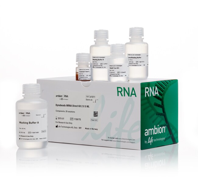
Kit de purificación Dynabeads™ mRNA DIRECT™
The Dynabeads™ mRNA DIRECT™ Kit is designed for simple and rapid isolation of pure, intact polyadenylated (polyA) mRNA directly from the crude lysate of animal and plant cells and tissues.
Have Questions?
Cambiar vista

| Número de catálogo | Cantidad |
|---|---|
| 61012 | 10 mL |
| 61011 | 5 mL |
Número de catálogo 61012
Precio (MXN)
-
Cantidad:
10 mL
El kit de ARNm DIRECT™ de Dynabeads™ está diseñado para el aislamiento rápido y sencillo de ARN mensajero (ARNm) poliadenilado puro e intacto (poliA) directamente de lisado crudo de tejidos y células animales y vegetales.El ARNm aislado es adecuado para su uso en todas las aplicaciones posteriores.Ventajas del kit de ARNm DIRECT™ de Dynabeads™:
•Rápido: un procedimiento de 15 minutos que produce ARNm puro intacto
•Aislamiento de ARNm de alta pureza: la mejor opción previa a la síntesis de ADN complementaria (ADNc)
•Sensibilidad del aislamiento de ARNm: permite la síntesis de ADNc y la construcción de bibliotecas de ADNc a partir de muestras de partida ultrapequeñas (permite la construcción de bibliotecas de ADNc a partir de una única célula)
Descripción general del sistema
El protocolo de aislamiento se basa en pares de base entre los residuos de poliA en el extremo 3' de la mayoría del ARNm y los residuos de oligo (dT)25 unidos covalentemente a la superficie del Dynabeads™.Otras especies de ARN que carecen de una cola poliA no se hibridan con los gránulos y se eliminan fácilmente.El ARN ribosómico, el ADN, las proteínas y las pequeñas moléculas de ARN (como ARN de transferencia, micro ARN y pequeños ARN nucleolares) no se unen a los gránulos y se descartan.Los agentes inhibidores de ARNasa en el tampón de lisis/unión junto con estrictas condiciones de lavado e hibridación garantizan el aislamiento de ARN puro intacto a partir de muestras crudas ricas en ARNasa, sin utilizar agentes caotrópicos fuertes.Los gránulos (beads) de purificación de ARNm se dirigen y capturan la transcriptoma de ARNm a partir de una amplísima variedad de muestras iniciales crudas (véase el protocolo).1 mg de gránulos Dynabeads™ oligo (dT)25 (200 μl) enlaza hasta 2 μg de ARNm.Una célula de mamífero típica contiene alrededor de entre 10 y 30 pg de ARN total, de los cuales entre un 1 % y un 5 % es ARNm.
Para uso exclusivo en investigación. No apto para uso en procedimientos diagnósticos.
Especificaciones
Volumen de elución10 to 100 μL
Tipo de producto finalARNm
Para utilizar con (aplicación)PCR en tiempo real cuantitativa (qPCR)
Para utilizar con (equipo)Automated Liquid Handling Systems
Características ecológicasBeads may be reused for multiple extractions
Compatibilidad de alto rendimientoCompatible con alto rendimiento
Cantidad10 mL
Condiciones de envíoTemperatura ambiente
Cantidad de material de partidaCells: ≤106
Plant: ≤400 mg
Tissue: ≤200 mg
Plant: ≤400 mg
Tissue: ≤200 mg
Producción2 μg mRNA per 200 μL of beads (Binding capacity)
Isolation TechnologyGránulo magnético
Tipo de muestraMuestras líquidas (p. ej., suero), muestras de plantas, muestras virales, células, tejido, muestras fijas y FFPE, levadura, ARN, sangre, muestras de plantas, muestras virales, células, tejido, muestras fijas y FFPE, sangre
Unit SizeEach
Contenido y almacenamiento
Contenido:
• 10 ml de Dynabeads con oligo (dT)25
• 60 ml de tampón de unión/lisis
• 120 ml de tampón de lavado A
• 60 ml de tampón de lavado B
• 15 ml de Tris-HCl de 10 mM (tampón de elución)
Contiene reactivos suficientes para 40 aislamientos estándar. Almacenar entre 2 °C y 8 °C.
• 10 ml de Dynabeads con oligo (dT)25
• 60 ml de tampón de unión/lisis
• 120 ml de tampón de lavado A
• 60 ml de tampón de lavado B
• 15 ml de Tris-HCl de 10 mM (tampón de elución)
Contiene reactivos suficientes para 40 aislamientos estándar. Almacenar entre 2 °C y 8 °C.
Preguntas frecuentes
I am getting DNA contamination after mRNA isolation using Dynabeads magnetic beads. Why is this?
My Dynabeads magnetic beads are not pelleting well with the magnet. Do you have any suggestions for me?
I have a long double-stranded DNA fragment I would like to isolate. What product do you recommend?
Can I use Dynabeads magnetic beads to isolate single-stranded DNA templates?
What is the magnetic susceptibility for Dynabeads magnetic beads?
Citations & References (6)
Citations & References
Abstract
Extracellular plasma RNA from colon cancer patients is confined in a vesicle-like structure and is mRNA-enriched.
Journal:RNA
PubMed ID:18456845
'Little is yet known about the origin and protective mechanism of free nucleic acids in plasma. We investigated the possibility of these free nucleic acids being particle associated. Plasma samples from colon cancer patients and cell culture media were subjected to various antibody incubations, ultracentrifugation, and RNA extraction protocols for
Relationships and differentially expressed genes among pancreatic cancers examined by large-scale serial analysis of gene expression.
Journal:Cancer Res
PubMed ID:11830538
'Pancreatic adenocarcinoma is among the most fatal of cancers, in part because of late diagnosis and a lack of effective therapies. Comprehensive studies are needed to better understand and address the cellular mechanisms and pathways of tumorigenesis. Serial analysis of gene expression was used to analyze gene expression profiles of
Stem cell and epithelial-mesenchymal transition markers are frequently overexpressed in circulating tumor cells of metastatic breast cancer patients.
Journal:Breast Cancer Res
PubMed ID:19589136
The persistence of circulating tumor cells (CTC) in breast cancer patients might be associated with stem cell like tumor cells which have been suggested to be the active source of metastatic spread in primary tumors. Furthermore, these cells also may undergo phenotypic changes, known as epithelial-mesenchymal transition (EMT), which allows
The transcription factors SOX9 and SOX10 are vitiligo autoantigens in autoimmune polyendocrine syndrome type I.
Journal:J Biol Chem
PubMed ID:11423552
Vitiligo is common in the hereditary disorder autoimmune polyendocrine syndrome type I (APS I). Patients with APS I are known to have high titer autoantibodies directed against various tissue-specific antigens. Using sera from APS I patients for immunoscreening of a cDNA library from human scalp, we identified the transcription factors
Disease-associated mutations in human mannose-binding lectin compromise oligomerization and activity of the final protein.
Journal:J Biol Chem
PubMed ID:14764589
Deficiency of human mannose-binding lectin (MBL) caused by mutations in the coding part of the MBL2 gene is associated with increased risk and severity of infections and autoimmunity. To study the biological consequences of MBL mutations, we expressed wild type MBL and mutated MBL in Chinese hamster ovary cells. The