Search


Citas y referencias (3)
Invitrogen™
SimplyBlue™ SafeStain
SimplyBlue SafeStain es una tinción Coomassie G-250 lista para usar, rápida, sensible y segura para visualizar las bandas de proteínasMás información
Have Questions?
Cambiar vista

| Número de catálogo | Cantidad |
|---|---|
| LC6065 | 3,5 L |
| LC6060 | 1 l |
Número de catálogo LC6065
Precio (MXN)
-
Cantidad:
3,5 L
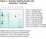
SimplyBlue SafeStain es una tinción Coomassie G-250 lista para usar, rápida, sensible y segura para visualizar las bandas de proteínas en geles de poliacrilamida y en membranas de PVDF secas. Es totalmente inocua y no requiere decolorantes ni fijadores de metanol ni ácido acético. El riesgo a una exposición peligrosa y los olores desagradables se han eliminado y se crea un entorno más saludable en el laboratorio.
Compare todas las tinciones de Coomassie ›
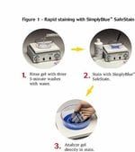
Protocolo fácil de usar
El protocolo de SimplyBlue SafeStain es fácil de seguir y puede finalizarse en menos de tres horas. Para mayor velocidad, un simple procedimiento de microondas se puede finalizar en 12 minutos. La decoloración no es necesaria, pero puede realizarse con agua desionizada para lograr la máxima sensibilidad, especialmente al realizar análisis posteriores, como la espectrometría de masas o cuando se requiere un fondo transparente.
Compare todas las tinciones de Coomassie ›
Protocolo fácil de usar
El protocolo de SimplyBlue SafeStain es fácil de seguir y puede finalizarse en menos de tres horas. Para mayor velocidad, un simple procedimiento de microondas se puede finalizar en 12 minutos. La decoloración no es necesaria, pero puede realizarse con agua desionizada para lograr la máxima sensibilidad, especialmente al realizar análisis posteriores, como la espectrometría de masas o cuando se requiere un fondo transparente.
Para uso exclusivo en investigación. No apto para uso en procedimientos diagnósticos.
Especificaciones
Ubicación de detecciónDetección en gel
Método de detecciónColorimétrico
Cantidad3,5 L
Duración de almacenamiento6 meses
Molécula dianaproteína
ColorBlue
Etiqueta o tinteCoomassie
Línea de productosSimplyBlue
Tipo de productoTinción segura de proteínas
Unit SizeEach
Contenido y almacenamiento
SimplyBlue™ SafeStain se suministra como reactivo de tinción listo para usar 1X. Almacenar a temperatura ambiente. Se garantiza la estabilidad durante 6 meses si se almacena correctamente. No hay cargos de HazMat asociados con este producto.
Preguntas frecuentes
Why is Coomassie G-250 used as the tracking dye in NuPAGE LDS Sample Buffer instead of bromophenol blue?
Is the SimplyBlue SafeStain the appropriate stain for quantitation by densitometry?
The sensitivity of my SimplyBlue SafeStain seems to be decreasing over time. Why is this?
How do I destain proteins on a PVDF membrane that were stained with SimplyBlue SafeStain?
How can membranes be stained?
Citations & References (3)
Citations & References
Abstract
2F3 monoclonal antibody recognizes the O26 O-antigen moiety of the lipopolysaccharide of enterohemorrhagic Escherichia coli strain 4276.
Journal:Clin Diagn Lab Immunol
PubMed ID:15138178
'Enterohemorrhagic Escherichia coli (EHEC) and enteropathogenic E. coli (EPEC) organisms are groups of pathogenic strains whose infections are characterized by a typical lesion of enterocyte attachment and effacement. They are involved in enteric diseases both in humans and in animals, and EHEC strains can be responsible for hemolytic uremic syndrome
Selective fluorescent labeling of S-nitrosothiols (S-FLOS): a novel method for studying S-nitrosation.
Journal:Nitric Oxide
PubMed ID:18706513
'Protein S-nitrosation is a reversible post-translation modification critical for redox-sensitive cell signaling that is typically studied using the Biotin Switch method. This method and subsequent modifications usually require avidin binding or Western blot analysis to detect biotin labeled proteins. We describe here a modification of the Biotin Switch assay that
Vascular endothelial growth factor-C and C-C chemokine receptor 7 in tumor cell-lymphatic cross-talk promote invasive phenotype.
Journal:Cancer Res
PubMed ID:19118020
'Most carcinomas spread to distant sites through lymphatic vessels. Several preclinical and clinical studies have shown a positive correlation between the incidence of lymph node metastasis and secretion of the lymphatic growth factor vascular endothelial growth factor-C (VEGF-C) by tumor cells, suggesting tumor lymphangiogenesis as an escape mechanism. However, recent