Search
Flow Cytometry Sample Preparation Buffers and Reagents

Flow cytometry buffers for sample preparation
Sample preparation is an important step for obtaining statistical value from flow cytometry experiments. Sample preparation reagents for flow cytometry include cell surface staining, intracellular and transcription factor staining buffer sets, cell lysis assays, blocking reagents, and magnetic cell isolation beads. Benefits of using these buffers include the following:
- Retains inherent biological characteristics
- Reduce background staining
- Quicker and easier preparation with optimized kits
Read the table below to select the buffer for your experiment.
Building a new panel and unsure of how to stain? Connect with your sales specialist to make a new panel and add a buffer
Flow cytometry buffer selection
| Cal-Lyse Solutions | High-Yield Lysis Solutions | ||
|---|---|---|---|
| Use |
Learn more about cell preservation and lysis buffers |
| |
| Contents |
|
| |
| Size | 25 mL | 100 mL | 500 mL |
| Cat. No. | GAS010 | GAS010S100 | HYL250 |
| Regulatory status | For research use only. | For research use only. | |
| When to use | Intended Use Statement: Intended to be used as an aid in the enumeration of leukocytes from various human biological sources including blood and bone marrow that have been stained with monoclonal antibodies for analysis using flow cytometric methods. | Intended Use Statement: Intended to be used to eliminate red cells from whole blood for flow cytometric analysis with minimal loss of rare blood cell populations. | |
| eBioscience Flow Cytometry Staining Buffer | |
|---|---|
| Use |
|
| Contents | Buffered saline solution:
|
| Size | 200 mL |
| Cat. No. | 00-4222-57 |
| eBioscience Intracellular Fixation & Permeabilization Buffer Set | FIX & PERM Cell Fixation & Cell Permeabilization Kit | |
|---|---|---|
| Use |
Learn more about the IC Fix & Perm buffer set |
|
| Contents |
|
|
| Size | 1 kit | 50 tests |
| Cat. No. | 88-8824-00 | GAS003 |
| Regulatory status | RUO – For Research Use Only. Not for Use in Diagnostic Procedures. | GPR – General Purpose Reagent. |
| When to use | For use in research only (i.e. research on any sample type). | For use in general laboratory application, that is used to collect, prepare, and examine specimens from the human body for diagnostic purposes. |
| eBioscience Foxp3 / Transcription Factor Staining Buffer Set | |
|---|---|
| Use |
|
| Contents |
|
| Size | 1 kit |
| Cat. No. | 00-5523-00 |
| eBioscience Super Bright Complete Staining Buffer | |
|---|---|
| Use |
|
| Contents |
|
| Size | 100 tests |
| Cat. No. | SB-4401-42 |
Cell isolation and expansion reagents
| Dynabeads for cell isolation | MagniSort Cell Separation Technology | |
|---|---|---|
| Use |
|
|
| Technology | Dynabeads activation and expansion technology mimics in vivo T cell activation. Available for human and mouse basic research as well as clinical research settings. Learn more | Starting with T cell–specific cocktails, use the MagniSort platform to obtain enriched T cells simpler, faster and with significant cost-savings compared to traditional column-based separation methods. Learn more |
Flow cytometry buffers for intracellular and cytoplasmic staining
Fixatives are necessary for saving samples to be used later or for looking at intracellular or intranuclear targets. Ready-to-use fixation kits are optimized for flow cytometry applications. Benefits of using these kits include the following:
- Methods used to stain cells take into consideration the location of the target proteins
- The fixation and permeabilization methods keep the morphological light-scattering characteristics of the cell intact
- Reagents in the kits help reduce background staining
Choosing the appropriate activating reagent will depend on (1) cell type, (2) expression level and kinetics of the protein of interest, and (3) experimental conditions.
Learn more about eBioscience Flow Cytometry Staining Buffer, Invitrogen FIX & PERM™ Cell Permeabilization Kit (RUO and GPR), Intracellular Fixation and Permeabilization Buffer Set (RUO), and the eBioscience Foxp3/ Transcription Buffer Set (RUO).

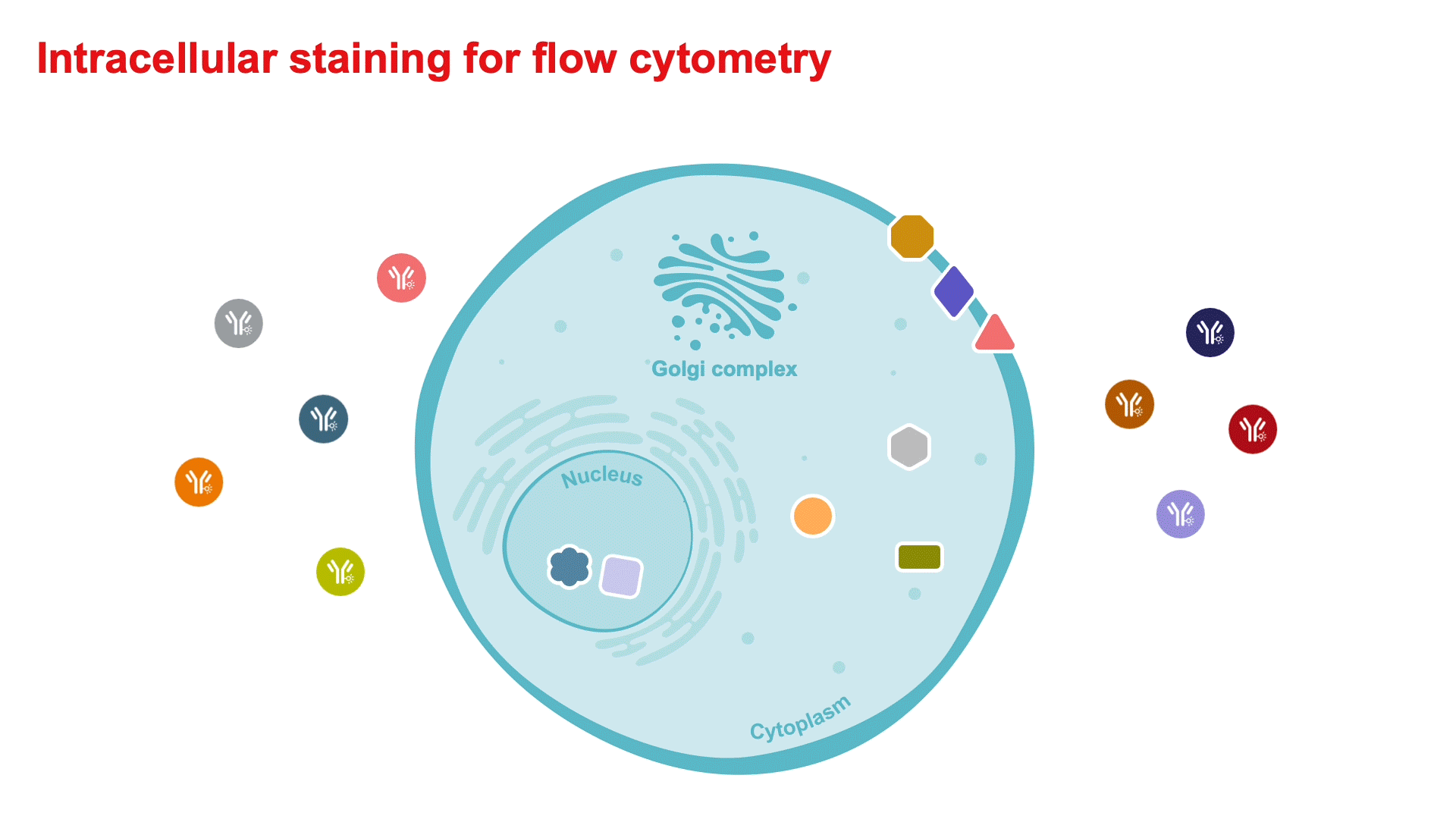
Figure 1. Cytoplasmic and intracellular staining procedure. The location of the target proteins within the cell is an important consideration in the selection of the appropriate buffer and staining protocol. For cytokine or phosphorylated proteins, timing of stimulants and treatment with protein transport inhibitors are required steps before staining.
Cytoplasmic and cytokine staining reagent kits
Activated cells often express higher levels of transcription factors, cytokines, chemokines, and other mediators detected by flow cytometry. Staining intracellular proteins for flow cytometry analysis often requires a fixation step to crosslink proteins and stabilize the cell membrane, as well as permeabilization to allow the antibodies access to the intracellular antigens. The Invitrogen eBioscience intracellular fixation and permeabilization buffers are designed for optimal detection of cytoplasmic and secreted proteins residing in organelles and vesicles. Benefits include:
- Compatible with a wide variety of markers and intracellular proteins
- Mild fixation and permeabilization for more accurate morphology
Learn more about eBioscience Flow Cytometry Staining Buffer and the Invitrogen FIX & PERM™ Cell Permeabilization Kit.

Figure 2. Staining of mouse CCL3 (MIP-1α) in macrophages. BALB/c thioglycolate-elicited peritoneal macrophages were unstimulated (left) or stimulated for 5 hours with LPS (Cat. No. 00-4976) (right) in the presence of Invitrogen eBioscience Protein Transport Inhibitor Cocktail (Cat. No. 00-4980). Cells were intracellularly stained with Anti–Mouse CD11b PerCP-Cyanine5.5 (Cat. No. 45-0112) and Anti–Mouse CCL3 (MIP-1α) PE (Cat. No. 12-7532) using the Invitrogen eBioscience Intracellular Fixation & Permeabilization Buffer Set (Cat. No. 88-8824) and protocol. Total viable cells, as determined by the Invitrogen eBioscience Fixable Viability Dye eFluor 450 (Cat. No. 65-0863), were used for analysis.
Nuclear staining reagent kits
Flow cytometry permits the detection of transcription factors within discrete immune cell subsets among a heterogeneous population and provides a sensitive approach to analyzing an immune response. Refer to the following section on intracellular staining buffers prior to any transcription factor staining analysis. Benefits include:
- Compatibility with all fluorophores including polymer dyes such as Brilliant Violets and Super Bright dyes.
- Lower signal-to-noise ratio, helping with low frequency signal.
- FoxP3 staining buffer has been tested on many antibodies targeting transcription factors and optimized for nuclear protein staining.

Figure 3. Staining of mouse E4BP4 (NFIL3) in stimulated NK cells. Mouse splenocytes unstimulated (left) or stimulated overnight with Invitrogen eBioscience Mouse IL-15/IL-15R Complex Carrier- Free Recombinant Protein (Cat. No. 34-8152 [right]) were stained with Anti–Mouse NK1.1 PE-Cyanine7 (Cat. No. 25-5941) and Anti–Mouse E4BP4 (NFIL3) PE (Cat. No. 12-5927). Intracellular staining for E4BP4 was performed using the Invitrogen eBioscience Foxp3/Transcription Factor Staining Buffer Set (Cat. No. 00-5523) and protocol. Cells in the lymphocyte gate were analyzed.
5 Steps to Intracellular Flow Cytometry
Discover products and resources that are available to help optimize your intracellular flow cytometry experiments.
Best practices for performing intracellular flow cytometry


Resources
Fluorophore and reagent selection guide for flow cytometry
Download Flow Cytometry Protocols Handbook
Spectral Flow Cytometry Fundamentals
Invitrogen eBioscience Resources—Selection guides, Best Protocols, product performance and more.
Intracellular Staining for Flow Cytometry How-To Video—for detecting cytokines and intranuclear markers.
Flow Cytometry Learning Center—Access flow cytometry educational resources for better experiment planning and execution.
Flow Cytometry Panel Builder—Design your flow cytometry panel with this online tool for a simplified, customizable experience to fit your needs.
5 Steps Resources
Support
Flow Cytometry Support Center—Find technical support recommendations for your flow cytometry workflows, including tips for experimental setup and in-depth troubleshooting help.
Flow Cytometry Panel Design Support—Work with one of our technical sales specialists to discuss your experimental needs and guide you through the process.
Not for resale. Super Bright Polymer Dyes are sold under license from Becton, Dickinson and Company.
*GPR—General Purpose Reagent.