Search
Invitrogen
TCR V alpha 3.2 Monoclonal Antibody (RR3-16), Brilliant Violet™ 421, eBioscience™
{{$productOrderCtrl.translations['antibody.pdp.commerceCard.promotion.promotions']}}
{{$productOrderCtrl.translations['antibody.pdp.commerceCard.promotion.viewpromo']}}
{{$productOrderCtrl.translations['antibody.pdp.commerceCard.promotion.promocode']}}: {{promo.promoCode}} {{promo.promoTitle}} {{promo.promoDescription}}. {{$productOrderCtrl.translations['antibody.pdp.commerceCard.promotion.learnmore']}}
Additional Information:
{{banner.description}}
")
FIGURE: 1 / 1
TCR V alpha 3.2 Antibody (404-5799-82) in Flow
BALB/c (left) and C57BL/6 (right) mouse lymph node cells were stained with CD3e Monoclonal Antibody, PE (Product # 12-0031-82) and 0.25 µg of V alpha 3.2 TCR Monoclonal Antibody, Brilliant Violet 421. Viable cells in the lymphocyte gate were used for analysis, as determined by 7-AAD (Product # 00-6993-50).

 in Flow")
Product Details
404-5799-82
Product Specifications
Species Reactivity
Mouse
Host/Isotype
Rat
/ IgG2b, kappa
Recommended Isotype Control
Class
Monoclonal
Type
Antibody
Clone
RR3-16
Conjugate
Excitation/Emission Max
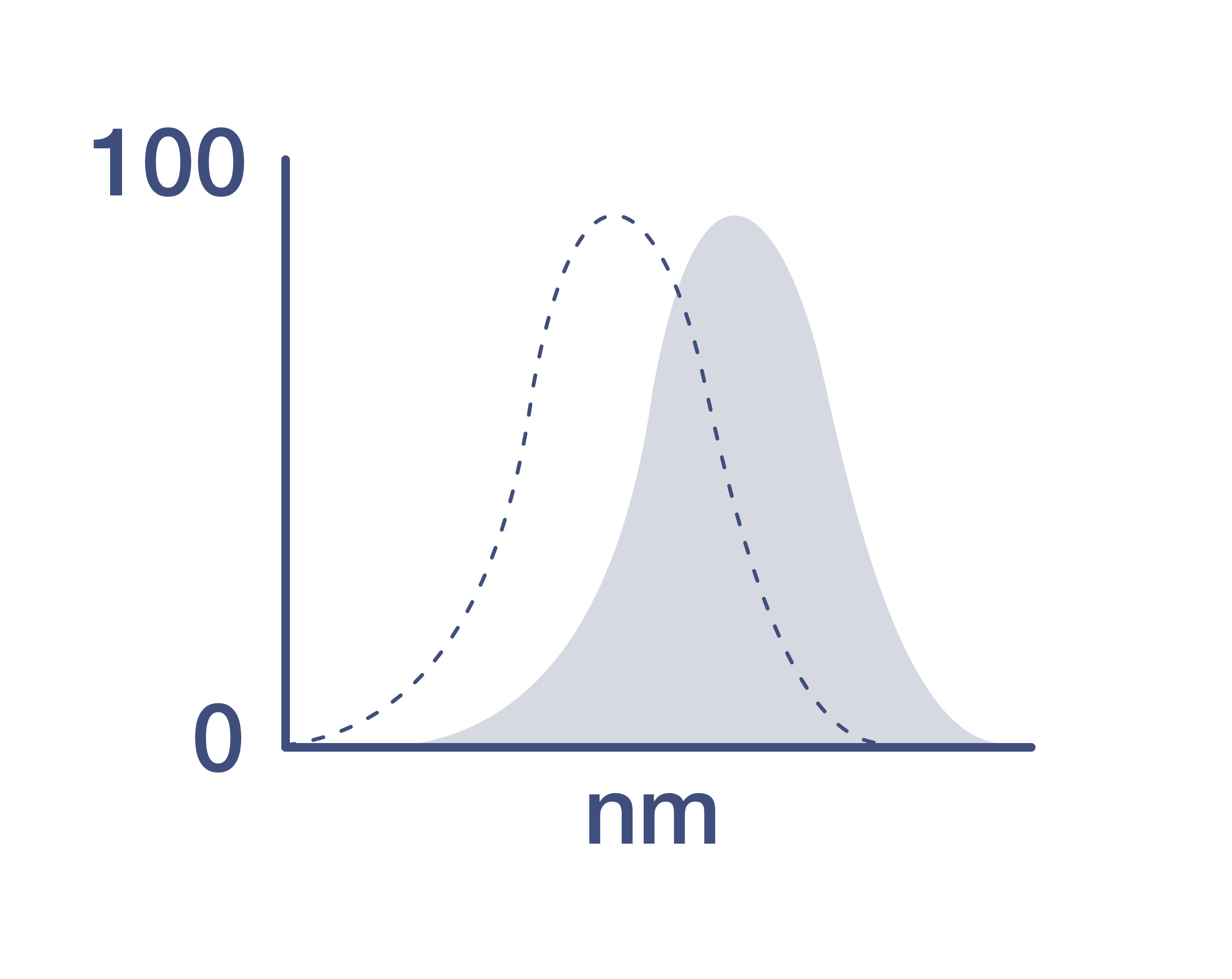
406/423 nm
View spectra
Form
liquid
Concentration
0.2 mg/mL
Amount
100 µg
Purification
Affinity chromatography
Storage buffer
PBS, pH 7.2, with BSA
Contains
0.09% sodium azide
Storage conditions
4°C, store in dark, DO NOT FREEZE!
Shipping conditions
Ambient (domestic); Wet ice (international)
RRID
Product Specific Information
Description:
This RR3-16 monoclonal antibody reacts with the mouse T cell receptor (TCR) V alpha 3.2 chain. Composed of an alpha and beta chain, TCR specificity is typically determined by Va, Ja, Vb, Db, and Jb gene rearrangement. The RR3-16 antibody recognizes the V alpha 3.2 chain on T cells from mouse strains bearing the b (e.g., C57BL/6) or c haplotype (e.g., SJL, SWR, and NOD) in the Tcra gene complex. The V alpha 3.2 chain is absent in mice with the a (e.g., Balb/c, AKR, C3H) and d (e.g., DBA/1 and DBA/2) haplotypes. Studies demonstrate that the V alpha 3.2 TCR is more highly expressed on CD8+ T cells.
Applications Tested:
This RR3-16 antibody has been tested by flow cytometric analysis of mouse lymph node cells. This may be used at less than or equal to 0.5 µg per test. A test is defined as the amount (µg) of antibody that will stain a cell sample in a final volume of 100 µL. Cell number should be determined empirically but can range from 10^5 to 10^8 cells/test. It is recommended that the antibody be carefully titrated for optimal performance in the assay of interest.
Blocking Buffers
When using two or more Super Bright, Brilliant Violet™, Brilliant Ultra Violet™, or other polymer dye-conjugated antibodies in a staining panel, it is recommended to use Super Bright Complete Staining Buffer (Product # SB-4401) or Brilliant Stain Buffer (Product # 00-4409-75) to minimize any non-specific polymer interactions. Please refer to the datasheet for Super Bright Staining Buffer or Brilliant Stain Buffer for more information.
Fixation
• Samples can be stored in IC Fixation Buffer (Product # 00-8222) (100 µL of cell sample + 100 µL of IC Fixation Buffer) or 1-step Fix/Lyse Solution (Product # 00-5333) for up to 3 days in the dark at 4°C with minimal impact on brightness and FRET efficiency/compensation.
• Some generalizations regarding fluorophore performance after fixation can be made, but clone specific performance should be determined empirically.
Excitation: 407 nm; Emission: 423 nm; Laser: Violet Laser.
BRILLIANT VIOLET™ is a trademark or registered trademark of Becton, Dickinson and Company or its affiliates, and is used under license. Powered by Sirigen™.
Target Information
The ability of T cell receptors (TCR) to discriminate foreign from self-peptides presented by major histocompatibility complex (MHC) class II molecules is essential for an effective adaptive immune response. TCR recognition of self-peptides has been linked to autoimmune disease. Mutant self-peptides have been associated with tumors. Engagement of TCRs by a family of bacterial toxins know as superantigens has been responsible for toxic shock syndrome. Autoantibodies to V beta segments of T cell receptors have been isolated from patients with rheumatoid arthritis (RA) and systemic lupus erythematosus (SLE). The autoantibodies block TH1-mediated inflammatory autodestructive reactions and are believed to be a method by which the immune system compensates for disease. Most human T cells express the TCR alpha-beta and either CD4 or CD8 molecule (single positive, SP). A small number of T cells lack both CD4 and CD8 (double negative, DN). Increased percentages of alpha-beta DN T cells have been identified in some autoimmune and immunodeficiency disorders. Gamma-delta T cells are primarily found within the epithelium. They show less TCR diversity and recognize antigens differently than alpha-beta T cells. Subsets of gamma-delta T cells have shown antitumor and immunoregulatory activity.
For Research Use Only. Not for use in diagnostic procedures. Not for resale without express authorization.
How to use the Panel Builder
Watch the video to learn how to use the Invitrogen Flow Cytometry Panel Builder to build your next flow cytometry panel in 5 easy steps.
References (0)
Have you cited this product in a publication?
Let us know so we can reference it here.
Bioinformatics
Protein Aliases: TCR V alpha3.2; TCRV alpha 3.2; TCRV alpha3.2; Va3.2; Valpha3.2
Disclaimer

Clicking the images or links will redirect you to a website hosted by BenchSci that provides third-party scientific content. Neither the content nor the BenchSci technology and processes for selection have been evaluated by us; we are providing them as-is and without warranty of any kind, including for use or application of the Thermo Fisher Scientific products presented.

Performance Guarantee
If an Invitrogen™ antibody doesn't perform as described on our website or datasheet,we'll replace the product at no cost to you, or provide you with a credit for a future purchase.*
Learn more
We're here to help
Get expert recommendations for common problems or connect directly with an on staff expert for technical assistance related to applications, equipment and general product use.
Contact tech support