100 kV cryo-TEM enables high-resolution single particle analysis
Within the past decade, cryo-electron microscopy (cryo-EM) techniques have revolutionized structural biology by enabling the visualization of challenging molecular and sub-cellular structures in their near-native states at high resolution. These valuable observations have, until recently, been relegated to advanced 300 kV instruments capable of rapid throughput and the highest contrast imaging. 200 kV microscopes have gained some traction for single particle analysis as well, but the ownership of either 200 or 300 kV cryo-transmission electron microscopes (cryo-TEMs) is still prohibitive for most labs, limiting the potential reach of cryo-EM. 100 kV instruments are more accessible, but have largely been regarded as sample screening tools rather than instruments capable of high-quality structural analysis.
A recent publication, led by scientists at Thermo Fisher Scientific, highlights novel advances in hardware that have changed this paradigm, enabling high-resolution structural analysis with the 100 kV Thermo Scientific Tundra Cryo-TEM, particularly of symmetrical molecules.

Bringing structural analysis to 100 kV cryo-TEM
Traditionally, structural analysis at 100 kV was primarily hampered by hardware limitations, from the electron source and optics down to the electron detector. To address this, the Tundra Cryo-TEM is equipped with a number of hardware improvements, such as an extreme brightness field emission gun (X-FEG), which is an electron source with a significantly lower energy spread that reduces the aberrations that can often occur at lower electron energies. Additionally, the arrangement of objective lenses in the Tundra Cryo-TEM produces a focal length of 2.3 mm, which greatly decreases chromatic aberrations, resulting in a >10x improvement in signal over former objective lens configurations with a 3.5 mm focal length. Finally, the type and pixel size of the electron detector inherently has a substantial impact on EM image quality. The Tundra Cryo-TEM is equipped with the Thermo Scientific Falcon C Direct Electron Detector, which offers a low signal-to-noise ratio and enhanced detective quantum efficiency (DQE) compared to scintillator-based detectors.
Together, these components provide a unique opportunity to perform single particle analysis at 100 kV. Through reduced aberrations and noise, high-quality images can routinely be obtained for 3D reconstruction.

Key molecules show potential of 100 kV cryo-EM
Eight benchmark proteins were analyzed with the Tundra Cryo-TEM using the Falcon C Detector. (An additional four samples were also analyzed with a scintillator-based Thermo Scientific Ceta Detector; more details can be found in the full publication.) Samples were categorized as either large (>200 kDa) and highly symmetric or smaller (<200 kDa) with lower symmetry. The Tundra Cryo-TEM was particularly successful at reconstructing the larger, highly symmetric proteins, consistently achieving <3 Å resolution.

The future of high-resolution cryo-EM at 100 kV
While 100 kV cryo-EM still has inherent limitations compared to 200-300 kV analysis, this promising research shows that structural analysis is indeed possible with these lower-energy microscopes. Instruments like the Tundra Cryo-TEM offer an accessible entry point into the world of cryo-EM structural analysis. No longer exclusively a tool for sample screening, these TEMs can produce high-resolution, informative structures for a variety of critical proteins.
We’re excited to contribute to the development of this technology and the increased access to single particle analysis that it will surely provide.
Learn more about the Tundra Cryo-TEM >>
3D Tissue Histology with Light-Sheet Microscopy Enables Nondestructive Analysis of Microglia
3D tissue analysis offers critical benefits for neuroscience... Alex Ilitchev, PhD
Read More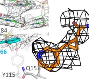
Fragment based drug discovery meets challenging drug targets with high-throughput cryo-EM
Benefits of FBDD in the search for novel therapeutics Frag... Dominic Meusch
Read More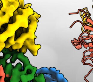
Targeted protein degradation as a novel therapeutic approach for undruggable diseases
Induced proximity for targeted protein degradation In 1993, ... Dominic Meusch
Read More
Analyzing Microparticles for Drug Delivery: the Critical Role of SEM and FIB-SEM Technology
Microparticles in drug development and delivery Microparticl...
Read More
Leave a Reply