With deeper insights into animal biology, researchers can better understand molecular function, leading not only to improved veterinary care, but also to the streamlined testing of new pharmaceutical agents that could aid in the design of new treatments for humans. The continued advancement of this work led Dr. Stephan Handschuh to a strong interest in 3D visualization and analysis of animal tissue images.
3D visualization and analysis at University of Veterinary Medicine, Vienna
A biologist at the University of Veterinary Medicine in Vienna, Handschuh works in the VetCore Facility for Research Imaging Unit where he supports researchers with their projects.
Austria’s only university for veterinary medicine, the University of Veterinary Medicine accommodates both an animal hospital and several research institutes. Researchers who use the VetCore Facility explore diverse fields ranging from animal physiology to cancer biology to veterinarian surgery.
 To accommodate this work, Handschuh helps researchers capture animal tissue images using a wide range of imaging techniques. He then often uses Thermo Scientific Amira Software to help researchers investigate all of their image data via 3D visualization and analysis.
“Amira includes virtually any function one could imagine when it comes to high-end image processing, segmentation, and visualization analysis,” says Handschuh. “This immensely broad functionality makes it quite unique.”
With Amira Software, those at the VetCore Facility can analyze the size and shape of organs, tissues, and cells. “We can visualize complex details of microscopic tissue anatomy, and we can put together data from different imaging modalities such as clinical CTs and MRIs and microCT, light microscopy, and electron microscopy—which deepens our understanding of biological structures,” says Handschuh.
His team uses hundreds of Amira Software features.
“This includes image segmentation, image co-registration of different data sets, advanced processing, image filtering, quantitative analysis, and of course all different kinds of visualization tools,” he adds.
A solution for complex cell biology imaging workflows
Since he started working at the university in 2012, Handschuh trained more than 100 students and post-doctorate researchers on how to use Amira Software.
He finds the ease at which his trainees have adopted the software fascinating.
“With just a couple hours of training, people who have never worked with this software really start to do this work on their own, and carry out quite complex tasks,” said Handschuh.

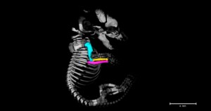
The software has been instrumental in analyzing skeletal development in mouse embryos, it’s been used to quantify movements in skull joints of miniaturized lizards, and it’s helped to identify approaches to brain surgery in cats and dogs.
“This functionality makes it possible to carry out complex workflows on a single software platform,” says Handschuh. “For us as facility staff supporting users, this brings clear advantages because users do not need to change between platforms during their analysis. This lowers the training effort and makes our lives much easier.”
“Furthermore, I love how Amira is very open,” he says. “It’s open with regard to integration of deep learning networks, but also for the integration of Matlab and Python, so this is also great.”
To learn more, please visit our Amira Software webpage.
Victoria Limosino is a marketing program manager at Thermo Fisher Scientific.


Leave a Reply