At the dawn of 2021, we introduced a new, more accessible solution for cryo-electron microscopy: the Thermo Scientific Tundra Cryo-TEM.

Jiri Novacek, PhD, Head of Core Facility, CEITEC using a new Tundra Cryo-TEM at Thermo Fisher Scientific in Brno
This smaller yet mighty cryo-EM instrument was designed to bring single particle analysis to every biochemistry laboratory. But what does that mean?
You asked. We answered.
Tundra Cryo-TEM: Commonly Asked Questions and Answers
What type of user and facility is the Tundra Cryo-TEM most suitable for?
The Tundra Cryo-TEM is an excellent choice for new users who are just getting started in cryo-EM. This is due to the guided workflow features and easy sample handling functions. It is also a useful addition for experienced users who are looking for an economical and flexible way to expand their cryo-EM sample preparation optimization capabilities.
Finally, even traditional room temperature microscopists may consider the Tundra Cryo-TEM to diversify their portfolio of services.

What are the dimensions of the Tundra Cryo-TEM?
Unlike other cryo-EM instruments, the Tundra Cryo-TEM is built to fit in most standard sized labs without any extensive modifications.
The entire system can fit into a 4 m X 4.2 m room with a ceiling height of at least 2.74 m.
This includes the microscope, support cabinets, sample loading station, HT tank and workstation.

What makes the Tundra Cryo-TEM suitable as a data collection tool, and not just a screening tool?
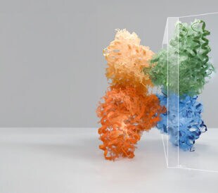
The Tundra Cryo-TEM is designed to deliver 3D reconstructions at biological relevant resolutions, particularly using the single particle method.
We have demonstrated this through a handful of examples, Gabaa receptor at 3.4A, T20S proteasome at 3.0A, Adeno-associated Virus 6 at 3.0A, and apoferritin as benchmark protein at 2.6A.
These amazing results are possible because the Tundra Cryo-TEM:
- Optimizes the optics for high resolution at 100kV.
- Has a CMOS detector. Not only does it have enhanced sensitivity and thus good signal-to-noise ratio at 100kV making it suited for extreme dose-sensitive samples in life science, but also it saves dose fractionations. Dose fractionations enable the teasing out of high-resolution information in the images that are otherwise blurred out by motion and radiation damage.
- Allows the sample in the column to be cooled by an extended liquid nitrogen Dewar that lasts 72-hours. This enables long data collection.

Download our infographic – Overcoming Misperceptions About Cryo-EM >>
What kind of camera is best for the Tundra Cryo-TEM and 100 kV applications?
Our Tundra Cryo-TEM is equipped with a scintillator-based CMOS camera, Ceta-F. Ceta-F has two major benefits, which makes it the best detector in the market for 100kV applications.
- Ceta-F has a very good Detective Quantum Efficiency (DQE) at 100kV. Traditionally scintillator-based cameras suffer from poor signal when detecting incoming electrons with high accelerating voltage, like 200kV or 300kV, because these electrons with high energy don’t interact with the scintillator too much. However, electrons at 100kV generate 2x more signal compared to 200kV.
- Ceta-F is radiation hard at 100kV. Direct Electron Detectors (DED) are superb in generating signal, but at the same time suffer from radiation damage. The damage is more severe with electrons at 100kV. For the similar reasons mentioned above, electrons at 100kV interact with the sensor more and causes more damage. None of the commonly known DEDs, such as Thermo Scientific Falcon, Gatan K3 and DE-20, have proven radiation hardness at 100kV.
What is the throughput for data collection on Tundra Cryo-TEM?
The throughput is 250-300 images/hour. The data sets we collected vary from 1,500 images (apoferritin, negative stain, or some other short screening sessions), to 5,000 images (proteins with symmetry), to 10,000 images (asymmetric protein).
Can we do multiple shots per hole on Tundra Cryo-TEM?
The Tundra Cryo-TEM has two condenser lenses and a mini-condenser lens. Having two condenser lenses means the illumination area is proportional to the C2 aperture.
We often use 50um and 70um C2 on the Tundra Cryo-TEM, which results in a 1.25um and 1.75um illumination area. With grid type R1.2/1.3, only one shot is possible on the Tundra Cryo-TEM. With grid type R2/2, when collecting close to the edge of the foil hole, it is possible to have multiple shots per hole. We have done two shots per hole with 5the 0um C2 aperture on R2/1 grid.
Can we use gold grids for data collection?
Yes, we can collect data with gold grids on the Tundra Cryo-TEM.
However, the challenge is with the automated coma-free alignment routine, which is desired before data collection. This cannot be performed on gold grids.
It is important to plan properly when working with gold grids. First, load a carbon grid and align the microscope to a perfect state, then load the gold grid for data collection.

How many grids can be screened on the Tundra Cryo-TEM in a day?
Ten grids can be screened on the Tundra Cryo-TEM per day. It takes 10 minutes to load a sample, 20 minutes to screen one grid, and another 10 minutes to unload.
It takes in total 40 minutes to screen one grid.
How often do I need to bakeout the sample loader and how long does it take?
When working under cryo conditions, the cryo loading station needs to be baked out at least daily. There are two bakeout modes:
- In-situ bakeout, which is fully automated, and takes eight hours to complete. This can be used when the microscope is running overnight.
- Ex-situ mode, in which user need to empty the liquid nitrogen pot manually. This takes roughly one hour and can be used during the day for a quick bakeout.
Are you doing demos, and can I send you samples?
Yes, we are doing demos and will accommodate standard samples for analysis!
Contact us to learn more and schedule a demo >>
View a special, 30-minute on-demand webinar for a closer look at our Tundra Cryo-TEM >>
//
Lingbo Yu is a product marketing manager at Thermo Fisher Scientific.
This post was written with the support of Dimple Karia, a product development engineer at Thermo Fisher Scientific.
Advances in high-resolution cryo-EM at 100 kV
100 kV cryo-TEM enables high-resolution single particle anal... Alex Ilitchev, PhD
Read More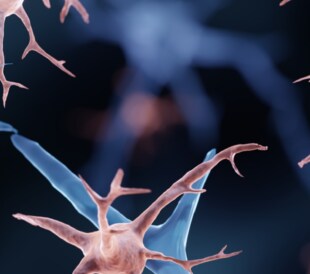
3D Tissue Histology with Light-Sheet Microscopy Enables Nondestructive Analysis of Microglia
3D tissue analysis offers critical benefits for neuroscience... Alex Ilitchev, PhD
Read More
Fragment based drug discovery meets challenging drug targets with high-throughput cryo-EM
Benefits of FBDD in the search for novel therapeutics Frag... Dominic Meusch
Read More
Targeted protein degradation as a novel therapeutic approach for undruggable diseases
Induced proximity for targeted protein degradation In 1993, ... Dominic Meusch
Read More
find beam error during alignment solution?